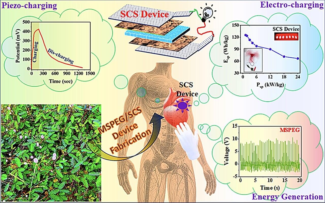
Most energy generators currently employed within the electronics industry are based on inorganic piezoelectric materials that are not bio-compatible and contribute to the pollution of the environment on Earth. In recent years, some electronics researchers and chemical engineers have thus been trying to develop alternative devices that can generate electricity for medical implants, wearable electronics, robots and other electronics harnessing organic materials that are safe, bio-compatible and non-toxic.
Researchers at the Materials Science Centre, Indian Institute of Technology Kharagpur recently introduced a new device based on seeds from the mimosa pudica plant, which can serve both as a bio-piezoelectric nanogenerator and a self-chargeable supercapacitor. Their proposed device, outlined in a paper published in the Chemical Engineering Journal, was found to achieve remarkable efficiencies, while also having a lesser adverse impact on the environment.
“This study was motivated by the need for biocompatible, self-sustaining energy systems to power implantable medical devices (e.g., pacemakers, neurostimulators) and wearable electronics,” Prof. Dr. Bhanu Bhusan Khatua, senior author of the paper, told Tech Xplore.
“Disembodied Brains: Understanding our Intuitions on Human-Animal Neuro-Chimeras and Human Brain Organoids” by John H. Evans Book Link: https://amzn.to/40SSifF “Introduction to Organoid Intelligence: Lecture Notes on Computer Science” by Daniel Szelogowski Book Link: https://amzn.to/3Eqzf4C “The Emerging Field of Human Neural Organoids, Transplants, and Chimeras: Science, Ethics, and Governance” by The National Academy of Sciences, Engineering and Medicine Book Link: https://amzn.to/4hLR1Oe (Affiliate links: If you use these links to buy something, I may earn a commission at no extra cost to you.) Playlist: • Two AI’s Discuss: The Quantum Physics… The hosts explore the ethical and scientific implications of brain organoids and synthetic biological intelligence (SBI). Several sources discuss the potential for consciousness and sentience in these systems, prompting debate on their moral status and the need for ethical guidelines in research. A key focus is determining at what point, if any, brain organoids or SBI merit moral consideration similar to that afforded to humans or animals, influencing research limitations and regulations. The texts also examine the use of brain organoids as a replacement for animal testing in research, highlighting the potential benefits and challenges of this approach. Finally, the development of “Organoid Intelligence” (OI), combining organoids with AI, is presented as a promising but ethically complex frontier in biocomputing. Our sources discuss several types of brain organoids, which are 3D tissue cultures derived from human pluripotent stem cells (hPSCs) that self-organize to model features of the developing human brain. Here’s a brief overview: • Cerebral Organoids: This term is often used interchangeably with “brain organoids”. They are designed to model the human neocortex and can exhibit complex brain activity. These organoids can replicate the development of the brain in-vitro up to the mid-fetal period. • Cortical Organoids: These are a type of brain organoid specifically intended to model the human neocortex. They are formed of a single type of tissue and represent one important brain region. They have been shown to develop nerve tracts with functional output. • Whole-brain Organoids: These organoids are not developed with a specific focus, like the forebrain or cerebellum. They show electrical activity very similar to that of preterm infant brains. • Region-specific Organoids: These are designed to model specific regions of the brain such as the forebrain, midbrain, or hypothalamus. For example, midbrain-specific organoids can contain functional dopaminergic and neuromelanin-producing neurons. • Optic Vesicle-containing Brain Organoids (OVB-organoids): These organoids develop bilateral optic vesicles, which are light sensitive, and contain cellular components of a developing optic vesicle, including primitive corneal epithelial and lens-like cells, retinal pigment epithelia, retinal progenitor cells, axon-like projections, and electrically active neuronal networks. • Brain Assembloids: These are created when organoids from different parts of the brain are placed next to each other, forming links. • Brainspheres/Cortical Spheroids: These are simpler models that primarily resemble the developing in-vivo human prenatal brain, and are particularly useful for studying the cortex. Unlike brain organoids, they do not typically represent multiple brain regions. • Mini-brains: This term has been debunked in favor of the more accurate “brain organoid”. These various types of brain organoids offer diverse models for studying brain development, function, and disease. Researchers are also working to improve these models by incorporating features like vascularization and sensory input. #BrainOrganoids #organoid #Bioethics #OrganoidIntelligence #WetwareComputing #Sentience #ArtificialConsciousness #Neuroethics #AI #Biocomputing #NeuralNetworks #ConsciousnessResearch #PrecautionaryPrinciple #AnimalTestingAlternatives #ResearchEthics #EmergingTechnology #skeptic #podcast #synopsis #books #bookreview #ai #artificialintelligence #booktube #aigenerated #documentary #alternativeviews #aideepdive #science #hiddenhistory #futurism #videoessay #ethics
European research led by University College London (UCL), together with Amsterdam UMC and the University of Basel, shows that a significant proportion of patients who suffer a stroke due to carotid artery narrowing can be treated with medication only.
A risky carotid artery operation, currently still the standard treatment for many patients, may then no longer be necessary for this group of patients. This research, published in The Lancet Neurology, may lead to the global guidelines for the treatment of these patients being adjusted.
In the Netherlands, about 2,000 people with carotid artery stenosis are operated on every year after they have had a stroke. Thirty years ago, large studies showed that an operation in which a narrowing in the carotid artery is removed reduced the risk of a new stroke.
The liver is the body’s control tower for metabolism, powering vital functions like converting nutrients to glucose, storing fat and breaking down toxins. Over a third of the world, however, is thought to be affected by conditions including metabolic dysfunction-associated steatotic liver disease (MASLD), which jeopardize key liver functions as the condition progresses. Hepatocyte organoids—the miniature, 3D models of the organ—hold immense promise for accelerating drug development and advancing regenerative therapies.
In a study published in Nature, Keio University researchers unveiled a method to proliferate these hard-to-grow organoids by a million-fold in just 3–4 weeks while maintaining key liver functions. “These organoids are potentially the closest laboratory representations of the liver and its multifunctionality,” says senior author Professor Toshiro Sato of the Keio University School of Medicine.
While organoids aim to mimic human organs, the liver’s repertoire of complex functions—and thus the energy it needs to operate—have made it challenging for researchers to grow organoids that proliferate and fully function, says Sato. When prioritizing growth and survival in laboratory settings, hepatocytes, the liver’s main cells, eventually transform into cells resembling cholangiocytes, which line the bile duct. Hepatocyte functions only last 1–2 weeks at most.
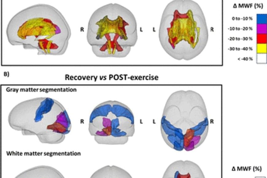
Exercise for a long period of time forces the human body to resort to its energy reserves. When running a marathon, for example, the body mainly consumes carbohydrates, such as glycogen, as a source of energy, but it resorts to fats when the glycogen in the muscles is used up. Myelin, which surrounds neurons in the brain and acts as an electrical insulator, mainly comprises lipids, and previous research in rodents suggests that these lipids can act as an energy reserve in extreme metabolic conditions.
A study conducted by researchers shows that people who run a marathon experience a decrease in the amount of myelin in certain regions of the brain. According to the study published by Nature Metabolism, this effect is completely reversed two months after the marathon.
The researchers used magnetic resonance imaging to obtain images of the brains of ten marathon runners (eight men and two women) before and 48 hours after the 42-kilometre race. Likewise, the researchers took images of the brains of two of the runners two weeks after the race, and of six runners two months after the race as a follow-up.
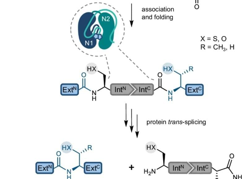
Proteins are the building blocks of life. They consist of folded peptide chains, which in turn are made up of a series of amino acids. From stabilising cell structure to catalysing chemical reactions, proteins have many functions. Their diversity is further increased by modifications that take place after the peptide chains have been synthesised. One form of modification is protein splicing. The protein initially contains a so-called ‘intein’, which removes itself from the peptide chain to ensure the correct folding and function of the final protein.
A research team has now answered a long-standing research question: Why does a special variant of the inteins, the ‘split inteins’, often encounter problems in the laboratory that significantly lower the efficiency of the reaction? The researchers were able to identify protein misfolding as one cause and have developed a method to prevent it.
The splicing of proteins rarely occurs in nature but is very interesting for research. The solution found by the team opens up possibilities for using split inteins to produce proteins that are useful in basic research or for applications in biotechnology and biomedicine.
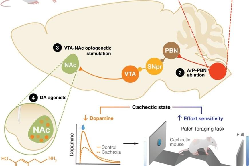
The fatigue and lack of motivation that many cancer patients experience near the end of life have been seen as the unavoidable consequences of their declining physical health and extreme weight loss. But new research challenges that long-held assumption, showing instead that these behavioral changes stem from specific inflammation-sensing neurons in the brain.
In a study published in Science, the researchers report that they identified a direct connection between cancer-related inflammation and the loss of motivation characteristic of advanced cancer. Studying mice with cancer-linked cachexia, a condition typical of the disease that leads to muscle wasting and weight loss, they discovered a previously unrecognized pathway in the brain. This pathway senses inflammation and actively suppresses dopamine — a key driver of motivation — resulting in apathy and loss of drive.
Blocking the pathway restored motivation, even though the cancer and weight loss continued. This indicates that apathy can be treated separately from the disease itself.