Podcast Episode · Your Brain On · 05/07/2025 · 52m


The new findings could help improve vaccine effectiveness in some immunocompromised patients. Researchers at the University of Colorado Anschutz Medical Campus have uncovered a critical, previously underappreciated role for B cells in vaccine protection. Best known for producing antibodies, B cells also guide other immune cells, specifically CD8 T cells, teaching them how to mount lasting defenses after vaccination.
The study was recently published in The Journal of Clinical Investigation.
“Think of CD8 T cells as rookie firefighters,” said lead author Jared Klarquist, PhD, assistant research professor of immunology and microbiology at the University of Colorado School of Medicine. “B cells teach the class on pacing. Without them, the rookies rush in, fight hard, and quit. They don’t save anything for the next fire.”
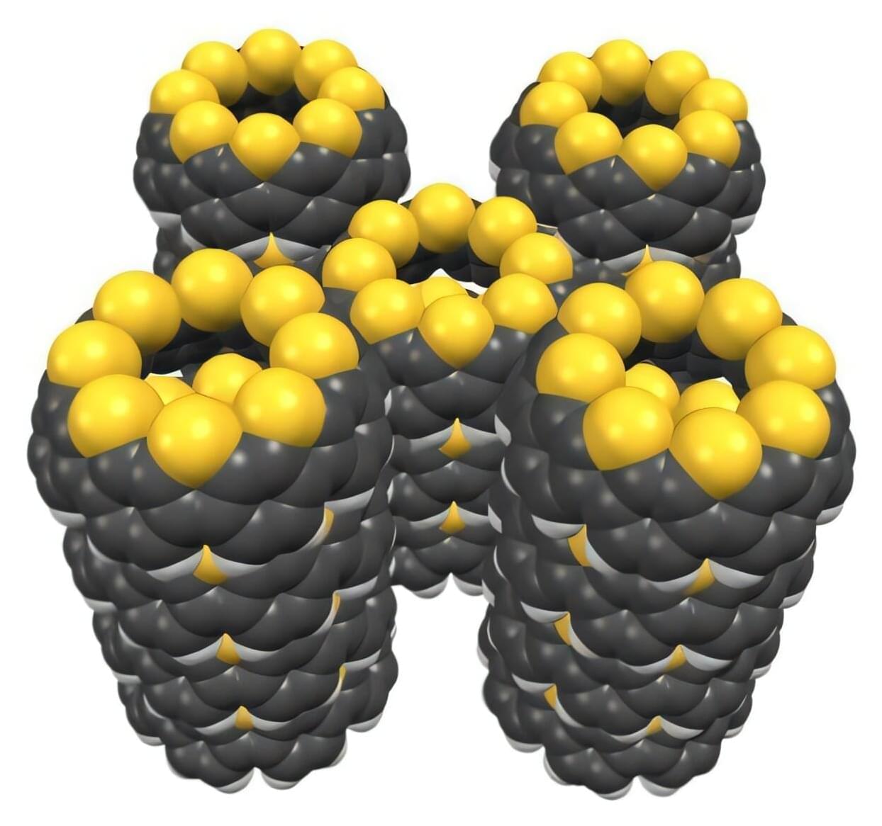
RIKEN chemists have hit upon a fast and easy way to combine so-called nanobelts of carbon with sulfur-containing functional groups. The work is published in the journal Nature Communications.
This new material has intriguing properties that make it promising for use in novel optoelectronic devices.
Ever since their discovery in 1991, carbon nanotubes—tiny hollow cylinders made entirely from carbon atoms—have been attracting a lot of interest, being used in applications ranging from electronics to medicine.

Introduction One thing newcomers to machine learning (ML) and many experienced practitioners often don’t realize is that ML doesn’t extrapolate. After training an ML model on compounds with µM potency, people frequently ask why none of the molecules they designed were predicted to have nM potency. If you’re new to drug discovery, 1nM = 0.001µM. A lower potency value is usually better. It’s important to remember that a model can only predict values within the range of the training set. If we’ve trained a model on compounds with IC50s between 5 and 100 µM, the model won’t be able to predict an IC50 of 0.1 µM. I’d like to illustrate this with a simple example. As always, all the code that accompanies this post is available on GitHub.
2024, for all of its challenges, has seen a remarkable amount of scientific discoveries by Israeli researchers across various disciplines.
From novel approaches to treating cancer to unraveling the intricacies of the human gut biome, these findings not only expand our understanding of the world but also pave the way for groundbreaking advancements in the future.
Let’s delve into 24 of the most fascinating discoveries made by Israeli scientists in 2024.
Take a look at these groundbreaking discoveries by Israeli researchers that are shaping our understanding of the world and its complexities.
Dr. Michael Osterholm, PhD, MPH ( https://www.cidrap.umn.edu/michael-t-osterholm-phd-mph ) is Regents Professor, McKnight Presidential Endowed Chair in Publi…
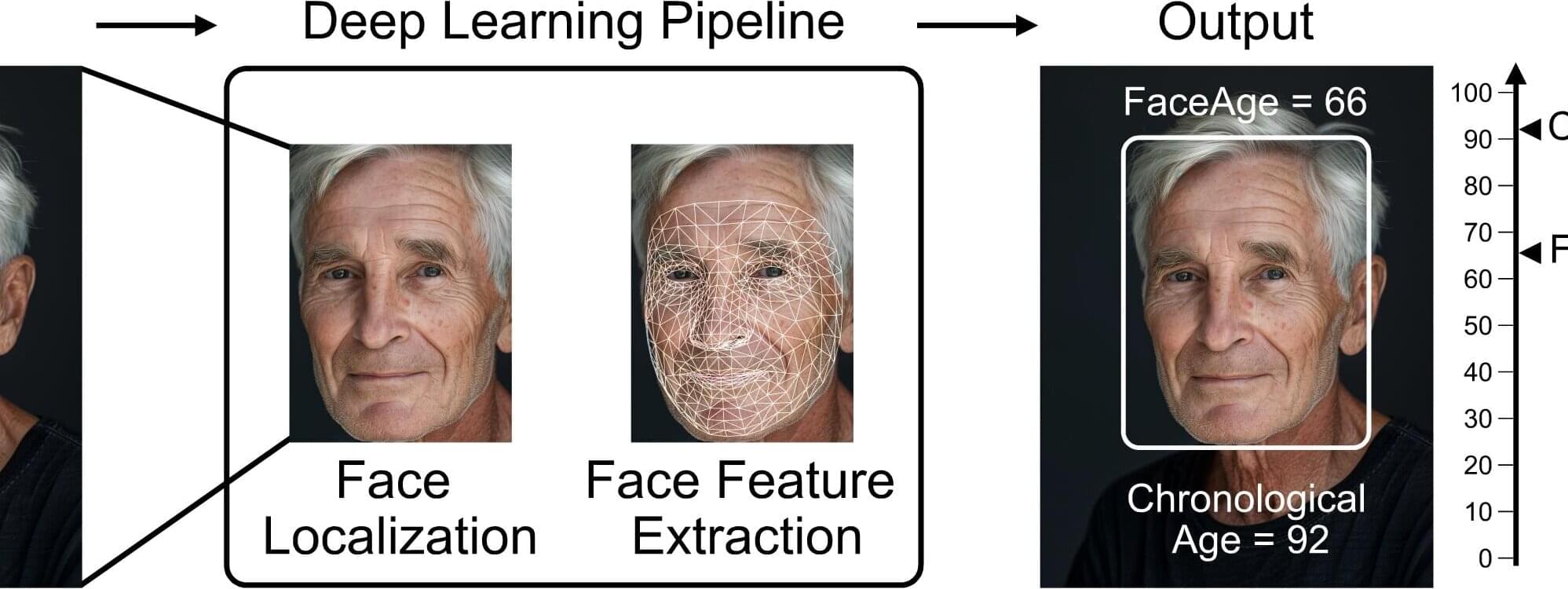
Eyes may be the window to the soul, but a person’s biological age could be reflected in their facial characteristics. Investigators from Mass General Brigham developed a deep learning algorithm called “FaceAge” that uses a photo of a person’s face to predict biological age and survival outcomes for patients with cancer.
They found that patients with cancer, on average, had a higher FaceAge than those without and appeared about five years older than their chronological age.
Older FaceAge predictions were associated with worse overall survival outcomes across multiple cancer types. They also found that FaceAge outperformed clinicians in predicting short-term life expectancies of patients receiving palliative radiotherapy.
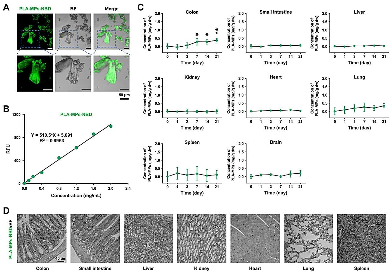
Microplastic pollution is a severe ecological and environmental issue and is also one of the important risk factors affecting human health. Polylactic acid (PLA), a medical biodegradable material approved by the FDA, is an important material to replace petroleum-based plastics.
Although PLA has achieved large-scale application in food packaging, its brittle characteristics make it more likely to generate microplastic particles. These particles can efficiently invade the gut through the food chain and trigger unknown biotransformation processes at the microbiota–host interface. Therefore, elucidating precisely the transformation map of PLA microplastics within the living body is crucial for assessing their safety.
In a study published in the Proceedings of the National Academy of Sciences, a research team led by Prof. Chen Chunying from the National Center for Nanoscience and Technology (NCNST) of the Chinese Academy of Sciences has revealed the complete biological fate of PLA microplastics (PLA-MPs) in the gut of mice, particularly focusing on their microbial fermentation into endogenous metabolites and their involvement in the carbon cycle.