The term resistant hypertension has been variably applied to patients with high blood pressure (BP) since at least the 1960s.1 Specific definitions have evolved over time to align with advances in treatment and BP goals. In 2018, the American Heart Association updated its definition of resistant…
Category: biotech/medical – Page 713

Artificial blood
Depending on the type of artificial blood that is made, various raw materials are used. Hemoglobin-based products can use either isolated hemoglobin or synthetically produced hemoglobin.
To produce hemoglobin synthetically, manufacturers use compounds known as amino acids. These are chemicals that plants and animals use to create the proteins that are essential for life. There are 20 naturally occurring amino acids that may be used to produce hemoglobin. All of the amino acid molecules share certain chemical characteristics. They are made up of an amino group, a carboxyl group, and a side chain. The nature of the side chain differentiates the various amino acids. Hemoglobin synthesis also requires a specific type of bacteria and all of the materials needed to incubate it. This includes warm water, molasses, glucose, acetic acid, alcohols, urea, and liquid ammonia.
For other types of hemoglobin-based artificial blood products, the hemoglobin is isolated from human blood. It is typically obtained from donated blood that has expired before it is used. Other sources of hemoglobin come from spent animal blood. This hemoglobin is slightly different from human hemoglobin and must be modified before being used.


Ingredient in energy drinks may help fuel aggressive blood cancers
A new study published in Nature has identified taurine, a common ingredient in energy drinks, as a nutrient that supports the survival and growth of leukemia stem cells in aggressive blood cancers.
Though taurine is known for its antioxidant and neuroprotective properties, the findings suggest it may play a harmful role in certain cancers.
The study’s authors caution that taurine supplements, including those in energy drinks, could influence disease progression in leukemia patients and recommend further research.


Advanced gene editor enables more precise insertion of complete genes
Ask scientists which gene-editing tool is most needed to advance gene therapy, and they’d probably describe a system that’s now close to realization in the labs of Samuel Sternberg at Columbia University Vagelos College of Physicians and Surgeons and David Liu at the Broad Institute of MIT and Harvard.
The gene editor—called evoCAST—goes a long way toward solving a problem that has confounded the development of gene therapies from the field’s beginnings: How to add long stretches of DNA to defined locations in the human genome without creating unwanted modifications.
The latest iteration of the editor, which utilizes complex enzymes found in bacteria, can be programmed to insert an entire gene—or multiple genes—into a specific location in the human genome with an efficiency suitable for gene therapy. Details of the editor are described in a paper published in Science.
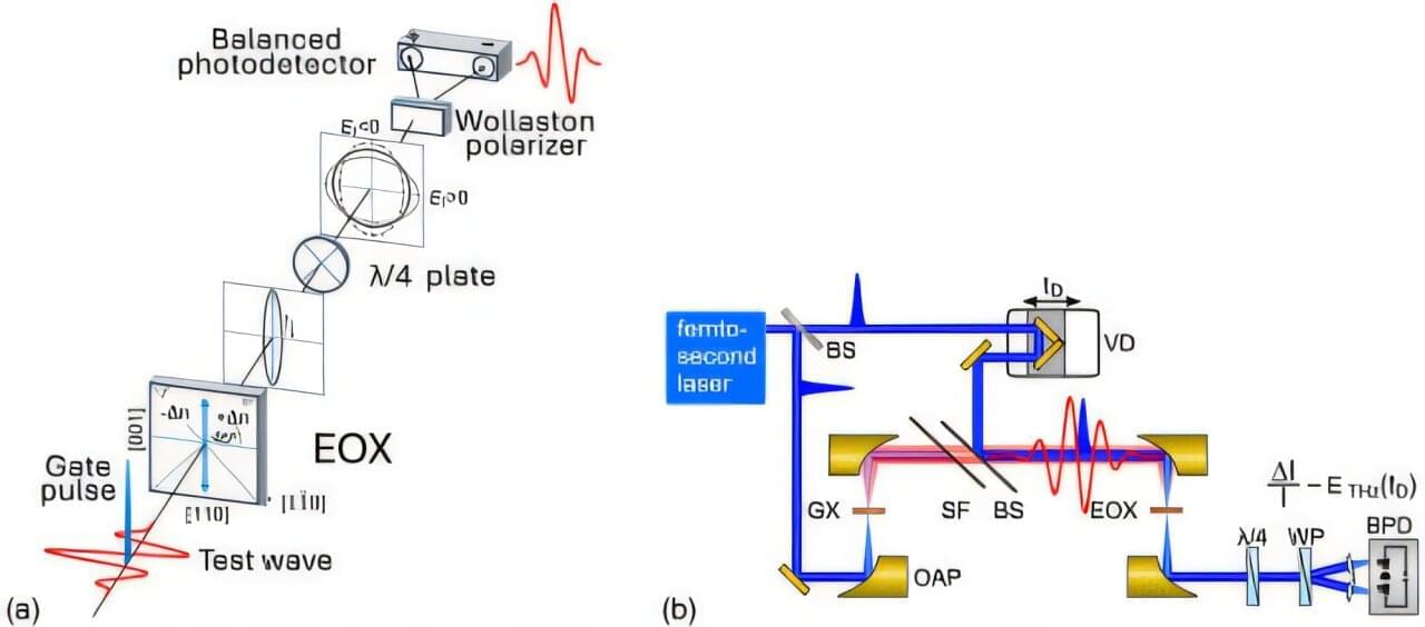
Electro-optic sampling research unlocks new insights into quantum physics
Konstantin Vodopyanov, a professor at the College of Sciences and CREOL, the College of Optics and Photonics, recently co-authored a study published in the journal Optica. This research examines electro-optic sampling (EOS), a technique that advances fields such as quantum physics, molecular spectroscopy and biomedical sensing.
As a professor at the two colleges, Vodopyanov shows how working across different fields can lead to new ideas. The Optica Fellow’s research, which combines interdisciplinary work, is shaping the future of quantum physics and other areas of science.
His new study explores how EOS transmits ultrashort laser pulses through crystals that change in response to an applied electric field. This technique allows researchers to accurately capture the shape and timing of electric fields across a broad range of frequencies.
Naturally occurring clay material has sought-after properties for use in quantum technology
In the future, quantum technology will become the standard for extremely fast computers. These kinds of machines will be important in everything from space technology to mineral exploration and the development of new medicines.
“Quantum technology is often associated with synthetic materials that have been developed in advanced, completely clean environments,” says Professor Jon Otto Fossum from NTNU’s Department of Physics.
But Fossum and colleagues have good news.