Scientists at St. Jude Children’s Research Hospital, the National Center for Genomic Analysis and the University of Adelaide have created a single-cell RNA analysis method that is 47 times cheaper and more scalable than other techniques.
Single-cell RNA sequencing provides scientists with important information about gene expression in health and disease. However, the technique is expensive and often prohibits analysis of large numbers of cells.
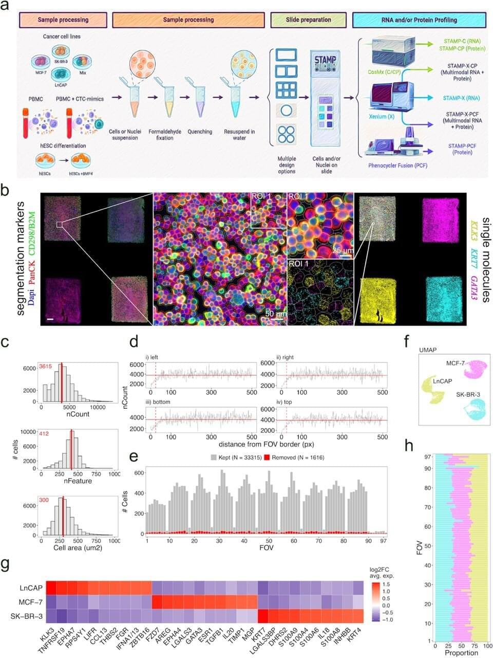
Scientists from St. Jude Children’s Research Hospital, the National Center for Genomic Analysis and the University of Adelaide have created a method that combines microscopy with single-cell RNA analysis to overcome these limitations. The technique called Single-Cell Transcriptomics Analysis and Multimodal Profiling through Imaging (STAMP) can look at millions of single cells for a fraction of the cost of existing approaches.