Two cannabis-derived compounds have shown remarkable effectiveness against fungal pathogens in laboratory tests, according to new Macquarie University research.
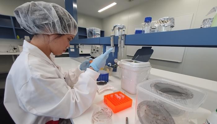
Promise: Dr Hue Dinh, pictured, and colleagues at the Macquarie University Galleria laboratory are hopeful their discovery will lead to new treatments for common skin infections.
In a study published in The Journal of Neglected Tropical Diseases (PLOS NTDs), researchers discovered that bioactives Cannabidiol (CBD) and Cannabidivarin (CBDV) killed harmful Cryptococcus neoformans - a WHO-listed priority fungal pathogen. The compounds also killed dermatophytes that cause common skin infections, and much faster than existing treatments.


