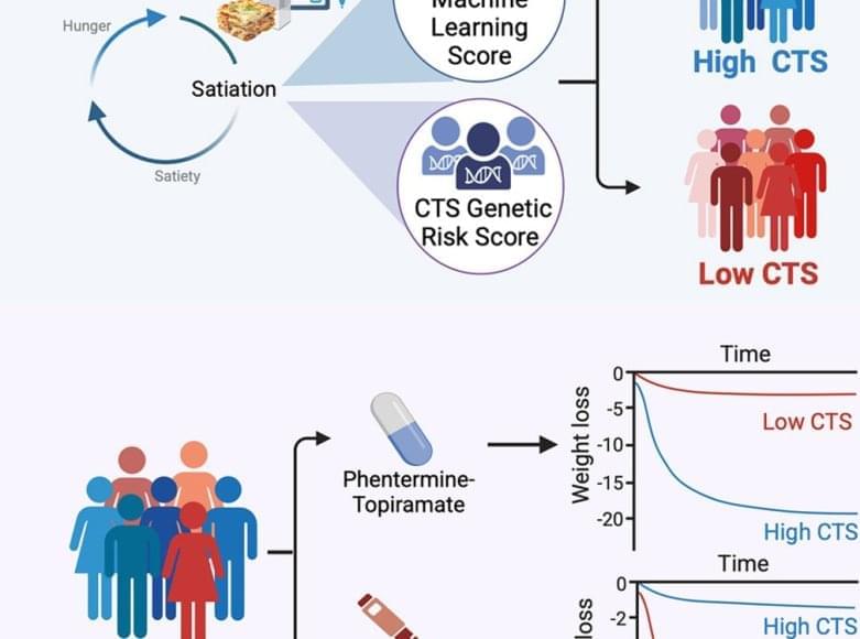
Large language models such as that used by ChatGPT could soon become essential tools for diagnosing and treating patients. To protect people’s privacy and safety, medical professionals, not commercial interests, must drive their development and deployment.
Category: biotech/medical – Page 534
Summary: Researchers developed ‘Anthrobots,’ microscopic biological robots made from human tracheal cells, demonstrating potential in healing and regenerative medicine.
These self-assembling multicellular robots, ranging from hair-width to pencil-point size, show remarkable healing effects, particularly in neuron growth across damaged areas in lab conditions.
Building on earlier Xenobot research, this study reveals that Anthrobots can be created from adult human cells without genetic modification, offering a new approach to patient-specific therapeutic tools.
The researchers are excited by the potential of how cells cooperate and communicate in the body and how they can be reprogrammed to create new structures and functions.
With the help of Simon Garnier at the New Jersey Institute of Technology, the team characterized the different types of Anthrobots that were produced.
They observed that bots fell into a few discrete categories of shape and movement, ranging in size from 30 to 500 micrometers (from the thickness of a human hair to the point of a sharpened pencil), filling an important niche between nanotechnology and larger engineered devices.
Some were spherical and fully covered in cilia, and some were irregular or football-shaped with more patchy coverage of cilia or just covered with cilia on one side. They traveled in straight lines, moved in tight circles, combined those movements, or just sat around and wiggled.
The accelerator, an advanced wakefield laser accelerator, is under 20 feet long, generating a 10 billion electron-volt (10 GeV) electron beam.
Bjorn “Manuel” Hegelich, associate professor of physics at UT and CEO of TAU Systems, alluding to the size of the chamber where the beam was produced stated: “We can now reach those energies in 10 centimeters.”
Scientists are aiming to use this technology for assessing the resilience of space-bound electronics against radiation, capturing the 3D internal configurations of emerging semiconductor chip designs, and potentially pioneering new cancer treatments and advanced medical imaging methodologies.
Furthermore, the statement noted that this accelerator could also be used to drive another device called an X-ray free electron laser, which could take slow-motion movies of processes on the atomic or molecular scale.
Summary: Researchers used AI to select and generate images for studying brain’s visual processing. Functional MRI (fMRI) recorded heightened brain activity in response to these images, surpassing control images.
The approach enabled tuning visual models to individual responses, enhancing the study of brain’s reaction to visual stimuli. This method, offering an unbiased, systematic view of visual processing, could revolutionize neuroscience and therapeutic approaches.
Are robots made from frog cells (Xenopus laevis).
Scientists have developed tiny robots made of human cells that are able to repair damaged neural tissue1. The ‘anthrobots’ were made using human tracheal cells and might, in future, be used in personalized medicine.
Developmental biologist Michael Levin at Tufts University in Medford, Massachusetts, and his colleagues had previously developed tiny robots using clumps of embryonic frog cells. But the medical applications of these ‘xenobots’ were limited, because they weren’t derived from human cells and because they had to be manually carved into the desired shape. The researchers have now developed self-assembling anthrobots and are investigating their therapeutic potential using human tissue grown in the laboratory. They published their findings in Advanced Science.
Levin and his team grew spheroids of human tracheal skin cells in a gel for two weeks, before removing the clusters and growing them for one week in a less viscous solution. This caused tiny hairs on the cells called cilia to move to the outside of the spheroids instead of the inside. These cilia acted as oars, and the researchers found that the resulting anthrobots — each containing a few hundred cells — often swam in one of several patterns. Some swam in straight lines, others swam in circles or arcs, and some moved chaotically.
The new contest aims to spur innovation into longer ‘healthspans’ with treatments that can actually reverse age-related degradation in body, mind and immune system.
Life Science Animation presents a groundbreaking 2D video animation that delves into the world of peptide-based cancer immunotherapies. In collaboration with Micropep, we explore Enterome’s OncoMimics platform, a novel approach to activating pre-existing memory T cell immunity for a more robust anti-tumor response.
Current immunotherapies often fall short as they only stimulate naïve T cell responses, leading to limited efficacy. Enterome’s innovative solution utilizes bacterial peptides, known as OncoMimics, that mimic tumor antigens. These OncoMimics are derived from common microbiome bacteria and are naturally tolerated by the human body.
In this video, we illustrate how OncoMimics cross the gut barrier, activating memory T cells and leading to a rapid and potent proliferation. The result? A targeted attack on tumor cells, leading to their destruction. Enterome’s OncoMimics are currently in human clinical development for the treatment of both solid and liquid tumors.
At Life Science Animation, we specialize in creating video animations that help explain complex scientific concepts, including those in biotech and aG tech. Our animations make science accessible and engaging for all audiences.
Science: Research suggest that dinosaurs 🦕 🦖 may have influenced how humans age today.
Human aging may have been influenced by millions of years of dinosaur domination according to a new theory from a leading aging expert. The ‘longevity bottleneck’ hypothesis has been proposed by Professor Joao Pedro de Magalhaes from the University of Birmingham in a new study published in BioEssays. The hypothesis connects the role that dinosaurs played over 100 million years with the aging process in mammals.
While some reptiles and amphibians show no significant signs of aging, all mammals—including humans—show a marked aging process.
Professor de Magalhaes’ hypothesis suggests that during the Mesozoic Era, mammals faced persistent pressure for rapid reproduction during the reign of dinosaurs, which over 100 million years led to the loss or inactivation of genes associated with long life, such as processes associated with tissue regeneration and DNA repair.
In a new study using brain scans of former NFL athletes, Johns Hopkins Medicine researchers say they found high levels of a repair protein present long after a traumatic brain injury such as a concussion takes place. The repair protein, known as 18 kDa translocator protein (TSPO), is known to be present in the brain at high levels in the immediate aftermath of brain injury as part of the inflammatory response and to facilitate repair. The new findings, published Oct. 30 in JAMA Network Open, suggest that brain injury and repair processes persist for years after players end collision sports careers, and lead to long-term cognitive problems such as memory loss.
“The findings show that participating in repeated collision sports like football may have a direct link to long-term inflammation in the brain,” says Jennifer Coughlin, M.D., associate professor of psychiatry and behavioral sciences at the Johns Hopkins University School of Medicine. Ongoing studies like the current one, she says, add details about how the brain heals — or doesn’t — and how repeated brain injuries, even mild ones that players routinely shake off, may over time affect cognitive abilities.
Want more breaking news? Subscribe for FREE to get the latest science stories delivered straight to your inbox. Don’t let the discoveries pass you by!