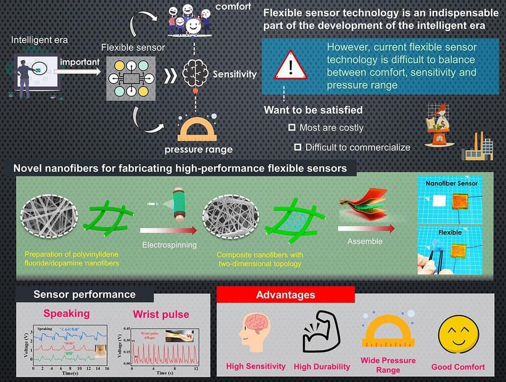
Toggle light / dark theme

Femoral head avascular necrosis (AVN) is a debilitating condition that prevents the thighbone from repairing itself at the portion closest to the hip, leading to possible collapse.

In a new study in Arthoplasty Today, a team including Yale Department of Orthopaedics & Rehabilitation’s Daniel Wiznia,…


In a paper published in the journal Arthroplasty Today, Daniel Wiznia, MD, assistant professor of orthopaedics & rehabilitation and co-director of Yale Medicine’s Avascular Necrosis Program, presents a new surgical technique designed to prevent or delay hip collapse in patients with femoral head avascular necrosis (AVN). Thanks to 3D innovations and novel applications of intraoperative navigation technology developed at Yale, Wiznia is leading a multidisciplinary approach to optimizing clinical outcomes.

Femoral AVN, otherwise known as osteonecrosis, is a debilitating condition associated with compromised blood supply to the portion of the thighbone closest to the hip. It particularly impacts the head of the bone. When the small vessels there are injured, the bone can no longer repair itself. Upwards of 20,000 new cases of femoral AVN are diagnosed each year in the United States, and those with the condition face a range of potential complications, such as collapse of the femoral head.

AVN is commonly diagnosed in people between the ages of 30 and 65. For some patients, there are no symptoms, which results in the condition being discovered incidentally. Up to 67 percent of patients with femoral AVN progress to symptomatic disease. A total hip arthroplasty (THA), otherwise known as a total hip replacement, is the current best treatment when the femoral head ultimately collapses. However, THA in younger patients has an increased risk of mechanical failure due to a higher level of physical activity and the length of time that the hip implant will be utilized. Therefore, there is a need for therapeutic strategies that effectively delay and prevent hip collapse, reducing the likelihood of requiring a THA.

There’s a lot to like about brain-computer interfaces, those sci-fi-sounding devices that jack into your skull and turn neural signals into software commands. Experimental BCIs help paralyzed people communicate, use the internet, and move prosthetic limbs. In recent years, the devices have even gone wireless. If mind-reading computers become part of everyday life, we’ll need doctors to install the tiny electrodes and transmitters that make them work. So if you have steady hands and don’t mind a little blood, being a BCI surgeon might be a job for you.

Shahram Majidi, a neurosurgeon at Mount Sinai Hospital in New York, began operating in clinical trials for a BCI called the Stentrode in 2022. (That’s “stent” as in a tube that often sits inside a vein or artery.) Here he talks about a not-too-distant future where he’s performing hundreds of similar procedures a year.

Brain-computer interfaces have been around for a few decades, and there are different kinds of implants now. Some have electrodes attached to your brain with wires sticking out of your head and connecting to a computer. I think that’s great as a proof of concept, but it requires an engineer sitting there and a big computer next to you all the time. You can’t just use it in your bedroom. The beauty of a BCI like the Stentrode, which is what I’ve worked with, is that nothing is sticking out of your brain. The electrodes are in blood vessels next to the brain, and you get there by going through the patient’s jugular. The receiver is underneath the skin in their chest and connected to a device that decodes the brain signals via Bluetooth. I think that’s the future.

A study led by Daniel Spratt, M.D., Vincent K. Smith Chair in Radiation Oncology at University Hospitals Seidman Cancer Center demonstrates the safety and efficacy of a novel oral hormone therapy, relugolix, in conjunction with radiation therapy for treating men with localized and advanced prostate cancer. This work is published in JAMA Oncology.

Only 3% to 5% of people who are diagnosed with this type of brain tumor will be alive three years later. On average, patients live about 14 months after diagnosis.

Now, an experimental therapy that reprograms a person’s own immune cells to attack these tumors is showing some exciting promise.

Three studies published within the past week have reported dramatic results with a therapy called CAR-T delivered directly to the brain. In some cases, tumors have seemingly melted away on brain scans by the next day.

A study of more than 21,000 average risk patients at 186 sites across the U.S., led by Regenstrief Institute and Indiana University School of Medicine research scientist Thomas Imperiale, M.D., has found that the next-generation multi-target stool DNA colorectal cancer screening test detects 94% of colorectal cancers. This test has the best performance for detection of both colorectal cancer and advanced precancerous polyps of any noninvasive colorectal cancer screening test.

The study results are published in the New England Journal of Medicine.

“We found that the next-generation stool DNA test had a good balance of sensitivity—detecting disease—and specificity—low false positive results. Compared to the fecal immunochemical test (FIT), the next gen test had superior sensitivity for both colorectal cancer and advanced pre-cancerous polys, especially the subgroup of advanced polyps containing high grade dysplasia,” said Dr. Imperiale, first author of the study.

A blood test for colon cancer performed well in a study published Wednesday, offering a new kind of screening for a leading cause of cancer deaths.

The test looks for DNA fragments shed by tumor cells and precancerous growths. It’s already for sale in the U.S. for $895, but has not been approved by the Food and Drug Administration and most insurers do not cover it. The maker of the test, Guardant Health, anticipates an FDA decision this year.

In the study, the test caught 83% of the cancers but very few of the precancerous growths found by colonoscopy, the gold standard for colon cancer screening. Besides spotting tumors, colonoscopies can prevent the disease by removing precancerous growths called polyps.

A newly developed “GPS nanoparticle” injected intravenously can home in on cancer cells to deliver a genetic punch to the protein implicated in tumor growth and spread, according to researchers from Penn State. They tested their approach in human cell lines and in mice to effectively knock down a cancer-causing gene, reporting that the technique may potentially offer a more precise and effective treatment for notoriously hard-to-treat basal-like breast cancers.