Chemotherapy, anti-cancer drugs that stop the growth of tumor cells, encompasses a wide variety of different agents. Some chemotherapeutic agents wor | Cancer

Made by U.S. pharmaceutical company Grail, the Galleri test aims to find fragments of DNA in a person’s blood that can indicate the presence of a cancerous tumor. Among the cancers that the test can detect, many have no current screening programs.
The PATHFINDER 2 study included more than 36,000 people aged 50 and older who had no cancer symptoms. In participants who were followed for more than a year, the test caught some 40.4% of cancer cases. For those who got a positive result on the Galleri test, 61.6% of them went on to be diagnosed with cancer—an improvement over previous trials of the test.
The results were presented on Saturday at the European Society for Medical Oncology meeting in Berlin, and have yet to be published in a peer-reviewed journal.

October 21–22, 2025 (Online) 🌿
Dear colleagues and friends.
We are pleased to invite you to the International Scientific Conference “Anti-Aging: Science and Practice of Healthy Longevity”, organized by the Gerontology Section of the Moscow Society of Naturalists (MOIP) at Lomonosov Moscow State University, with the support of the Gerontology Society of the Ural Branch of the Russian Academy of Sciences (URAN).
📅 Dates: October 21–22, 2025 🕛 Time: 12:00–16:00 (Moscow time) 💻 Format: Online participation (free of charge) 🗣️ Working language: Russian.
🔹 October 21 — “Hypoxic Training (Therapy): Modern Aspects of Healthy Longevity Medicine” 🔹 October 22 — “Fundamental and Clinical Gerontology as the Basis of Healthy Longevity Medicine”
The conference will feature leading scientists from Russia, Germany, Belarus, Kyrgyzstan and other countries. Topics include: • Hypoxic therapy and adaptive mechanisms; • Geroprotection and the biology of aging; • Epigenetic reprogramming and cellular rejuvenation; • Applied aspects of active and healthy longevity.
🔗 Connection links: • Day 1 (October 21): https://my.mts-link.ru/j/38630705/5798697072
The gang catches up with Emil Kendziorra after the Biostasis 2025 conference at the European Biostasis Foundation. Watch it on YouTube here. Topics covered include:
• How to get a Tomorrow Bio ambulance in your hometown.
• Tomorrow Bio’s plan to collect brain samples to check ultra-structure preservation in its cryonics patients — and how it will respond to what it finds.
• What’s new and what’s next for Tomorrow Bio.
• Our near death experiences.
Links:
• Cryosphere Discord Server: / discord.
• Cryonics Subreddit: / cryonics.
As Parkinson’s disease progresses, harmful protein clumps build up in the brain, blocking communications between neurons and killing them off – but what if we could prevent these clusters from forming?
Researchers led by a team from the University of Bath in the UK have achieved just that in a basic worm model of Parkinson’s. They engineered a peptide, a small amino acid chain, to essentially keep a protein called alpha-synuclein locked in its healthy shape. This prevented the misfolding that leads to clumps.
The potential treatment checks several important boxes: it’s durable, and it can survive inside cells without causing any toxic side effects.
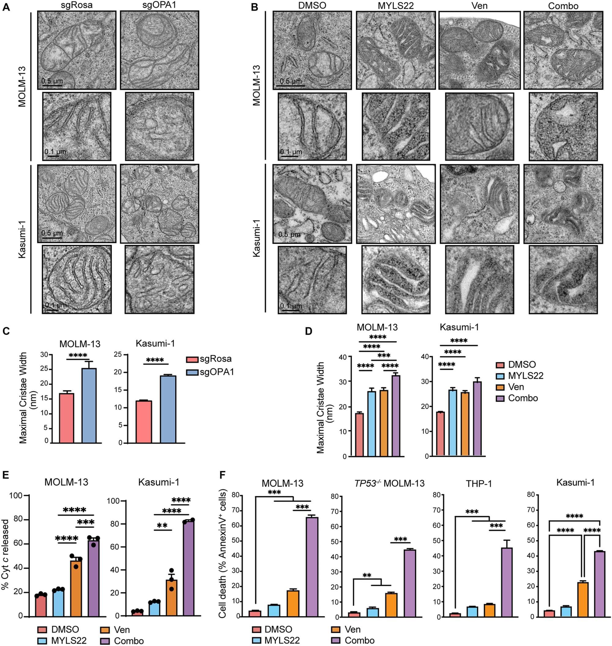
Researchers from Rutgers Health and other institutions have discovered why a powerful leukemia drug eventually fails in most patients—and found a potential way to overcome that resistance.
Team members identified a protein that lets cancer cells reshape their energy-producing mitochondria in ways that protect them from venetoclax (brand name, Venclexta), a standard treatment for acute myeloid leukemia that often loses effectiveness after prolonged use.
Blocking that protein with experimental compounds in mice with human acute myeloid leukemia restored the drug’s effectiveness and prolonged survival.
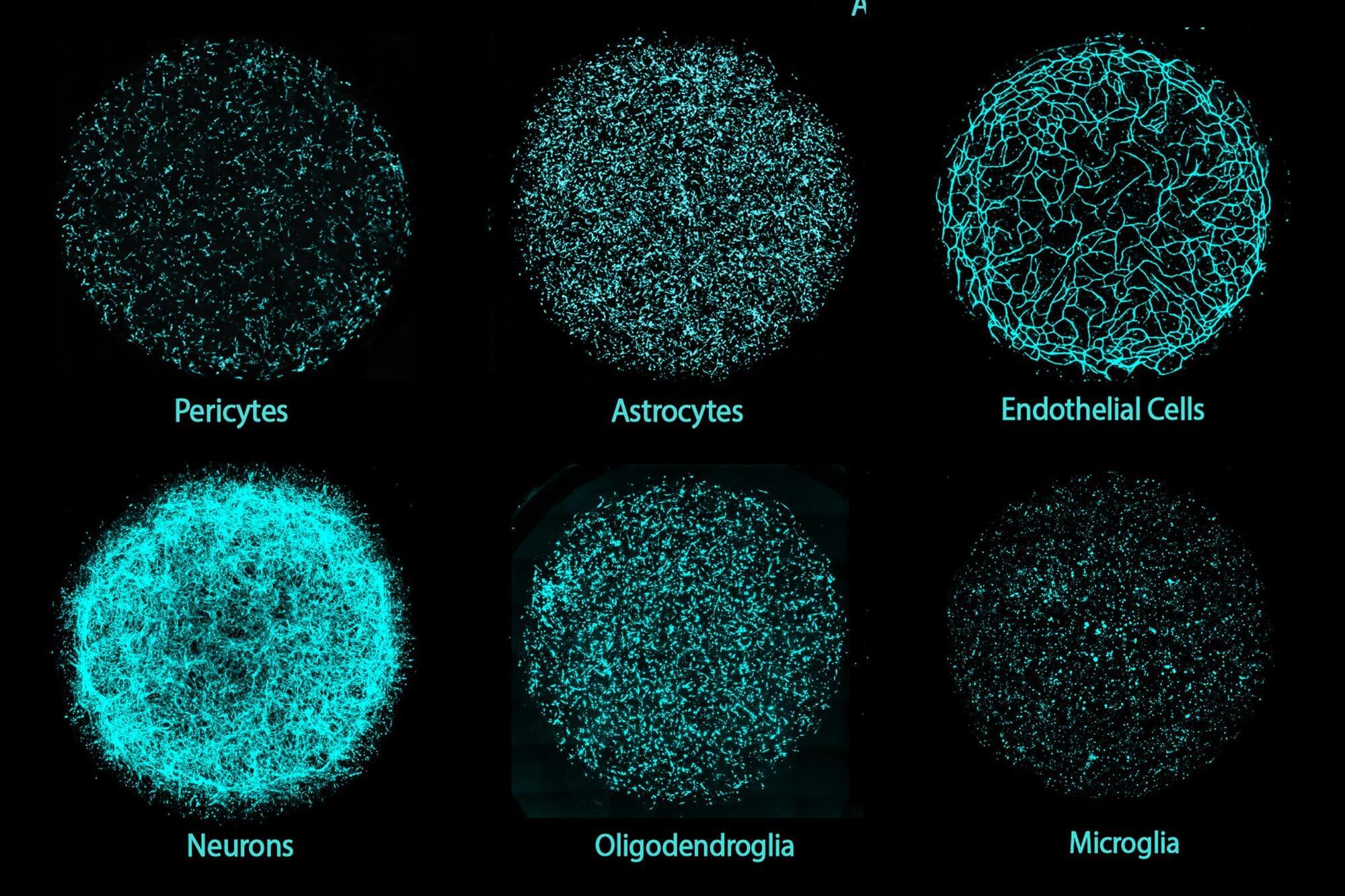
A new 3D human brain tissue platform developed by MIT researchers is the first to integrate all major brain cell types, including neurons, glial cells and the vasculature into a single culture. Grown from individual donors’ induced pluripotent stem cells, these models—dubbed Multicellular Integrated Brains (miBrains)—replicate key features and functions of human brain tissue, are readily customizable through gene editing, and can be produced in quantities that support large-scale research.
Although each unit is smaller than a dime, miBrains may be worth a great deal to researchers and drug developers who need more complex living lab models to better understand brain biology and treat diseases.
“The miBrain is the only in vitro system that contains all six major cell types that are present in the human brain,” said Li-Huei Tsai, Picower Professor, director of The Picower Professor of Learning and Memory, and senior author of the study describing miBrains, published in the Proceedings of the National Academy of Sciences.
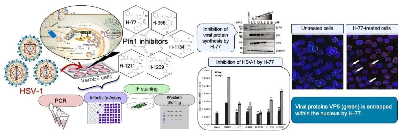
A class of antivirals called Pin1 inhibitors could reduce or stop outbreaks of herpes simplex virus 1 (HSV-1), the common infection behind oral herpes, according to new research published in Antiviral Research.
HSV-1 causes sores around the mouth, commonly called cold sores or fever blisters. Most people are infected with HSV-1 in childhood, and between 50% and 90% of people worldwide have HSV-1. After the initial infection, HSV-1 remains in the body and can reactivate throughout a person’s life. While HSV-1 infections are usually mild, they can be serious and even deadly for people with suppressed immune systems. Finding new, more effective antivirals for this common illness is essential.
Researchers focused on an enzyme called peptidyl-prolyl cis-trans isomerase NIMA-interacting 1, or Pin1, that regulates protein stability, function, and cellular structure. When this enzyme is dysregulated, it can play a role in a variety of conditions, including obesity, cancer, heart failure, and more. Viruses, such as cytomegalovirus (CMV) and SARS-CoV-2, are known to affect Pin1, and Pin1 inhibitors have been developed to reduce the impact of these viruses.

Clinical resistance is a complex phenomenon in major human cancers involving multifactorial mechanisms, and hypoxia is one of the key components that affect the cellular expression program and lead to therapy resistance. The present study aimed to summarize the role of hypoxia in cancer therapy by regulating the tumor microenvironment (TME) and to highlight the potential of hypoxia-targeted therapy.