Flatworms can rebuild themselves from just a small fragment, and now scientists know why. Their stem cells ignore nearby instructions and respond to long-distance signals from other tissues. This discovery turns old stem cell theories upside down and could lead to new ways to repair or regrow human tissue. It also reveals a hidden complexity in one of nature’s simplest creatures.
Category: biotech/medical – Page 475
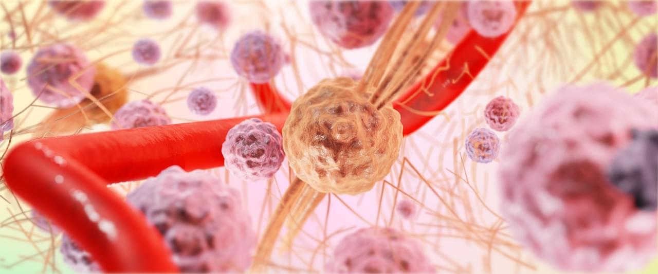
Engineered Immune Cells Improves Anti-Cancer Response
Scientists have developed a way to engineer immune cells that specifically target tumors. The application of engineering cells first appeared in the 1980s, but the concept has significantly progressed over the last few decades. This approach of engineering a patient’s cells as a form of therapy allow the immune system to specifically target the tumor and limit off-target affects.
Chimeric antigen receptor (CAR) T cells is an immunotherapy that takes patient T cells and edits them to target the tumor. The cells are then reinfused to accurately and effectively eliminate tumor growth. Immunotherapy is a general classification of cancer treatments that refers to the redirection of the immune system toward a disease or infection. T cells are responsible for the identification and elimination of infected cells and other diseases. Therefore, they are the optimal cell to engineer for robust and durable antitumor immunity. While scientists are working to engineer other cell types, CAR T cell therapy have been shown to have improved efficacy in multiple types of blood or hematological malignancies.
CAR T cell therapy in solid tumors is less effective. Unfortunately, the environment around the tumor has a complex network of various cell types combined with proteins and other molecules that inhibit CAR T cell efficacy. As a result, these CAR T cells cannot function and contribute to tumor progression. Scientists are currently working to improve CAR T cell therapy and develop stronger anti-cancer treatments.

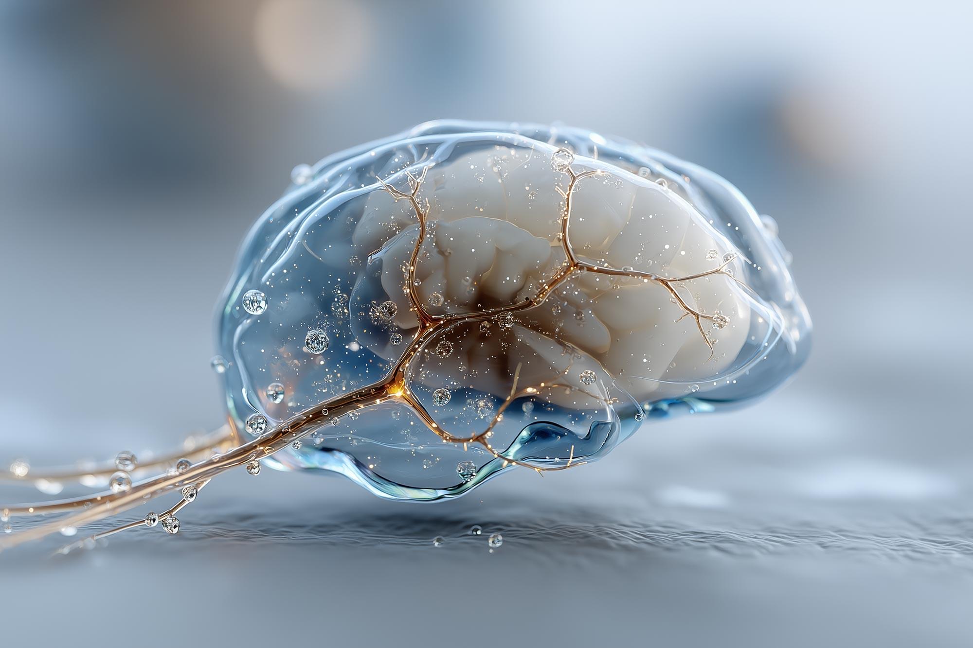
Previously unrecognized hub in the brain’s lymphatic drainage system may assist with clearing waste
How does the brain take out its trash? That is the job of the brain’s lymphatic drainage system, and efforts to understand how it works have pushed the boundaries of brain-imaging technologies.
A new study in iScience by researchers at the Medical University of South Carolina reveals—for the first time in humans—evidence of a previously unrecognized hub in the brain’s lymphatic drainage system—the middle meningeal artery (MMA).
Taking advantage of a NASA partnership that provided access to real-time MRI technologies originally developed to study how spaceflight affects fluid dynamics in the human brain, the MUSC research team, led by Onder Albayram, Ph.D., tracked cerebrospinal and interstitial fluid flow along the MMA in five healthy participants over a six-hour period. They found that the drainage flow of the cerebrospinal fluid was passive, suggesting lymphatic rather than blood flow. Blood would have had a faster, more dynamic flow.
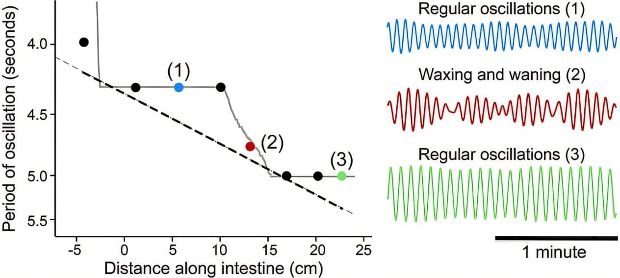
How a chorus of synchronized frequencies helps you digest your food
Synchronization abounds in nature: from the flashing lights of fireflies to the movement of fish wriggling through the ocean, biological systems are often in rhythmic movement with each other. The mechanics of how this synchronization happens are complex.
For instance, in the vasculature of the brain, blood vessels oscillate, expanding and contracting as needed. When there is neural activity, the arterioles expand to increase blood flow, oxygen and nutrients. These oscillations are self-sustained, but the arterioles also work in concert with each other. How this happens is not well understood.
To uncover the answer, researchers at the University of California San Diego looked to another part of the body: the gut. Here they found that oscillators operating at similar frequencies lock onto each other in succession, creating a staircase effect. Their work appears in Physical Review Letters.
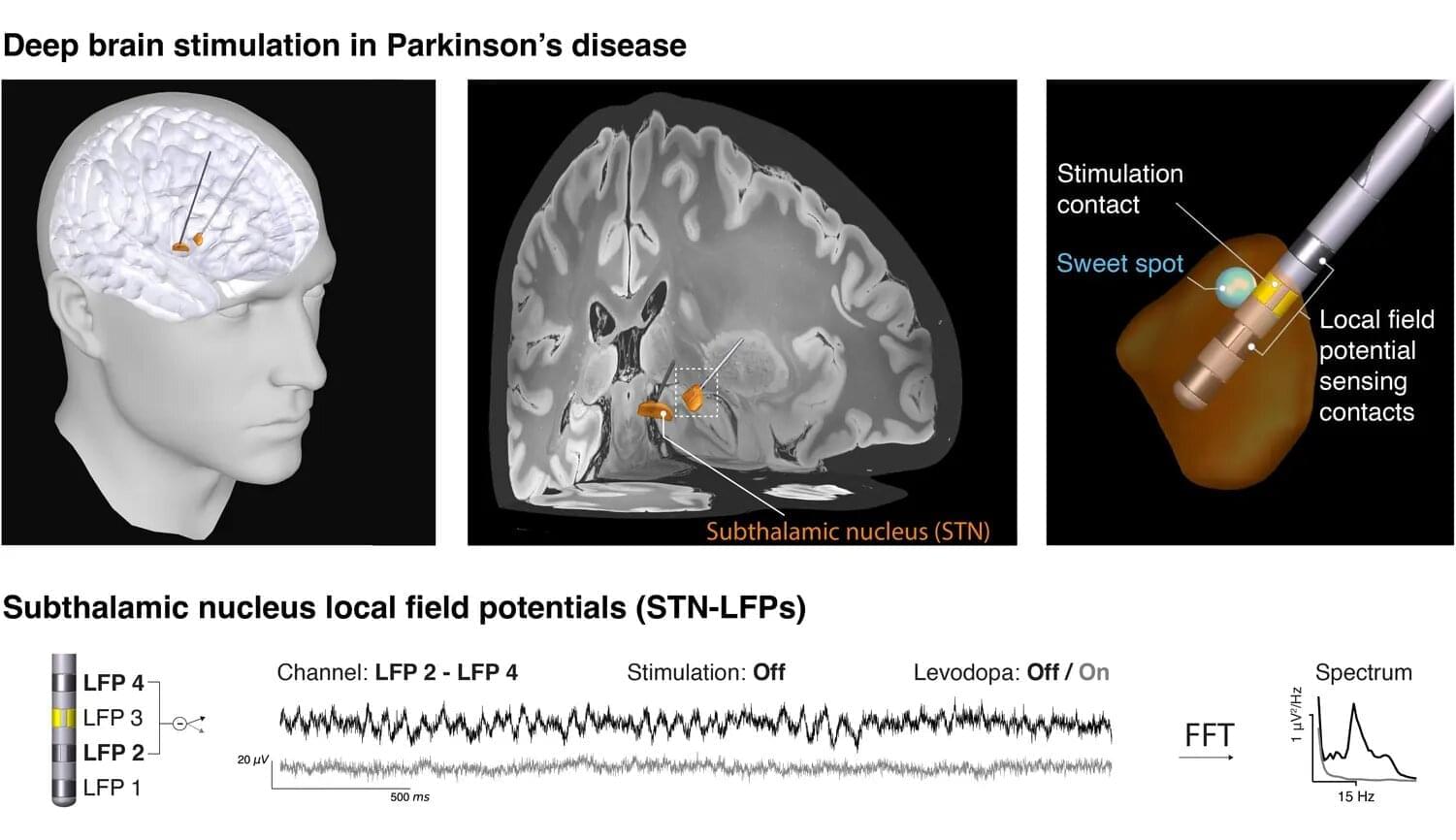
New electrical signature of Parkinson’s disease identified
What happens in the brain when a person experiences the characteristic movement symptoms of Parkinson’s disease? Researchers around the world are seeking answers through various approaches. One of these builds on a treatment already established in clinical care: deep brain stimulation. In this therapy, stimulating electrodes are implanted in patients’ brains to alleviate symptoms using electrical impulses. The same electrodes also enable unique electrical measurements from areas otherwise inaccessible in humans. These data can help uncover the neural mechanisms of Parkinson’s disease and inspire new therapeutic strategies.
In close collaboration with leading European deep brain stimulation centers—including Charité Berlin, Heinrich-Heine University Düsseldorf, University College London, and the University of Oxford—the Max Planck team has now taken an important step forward. For their study, now published in eBioMedicine, the researchers focused on so-called “beta waves,” which oscillate ca. 20 times per second and whose strength is thought to correlate with the severity of movement symptoms.
However, when reviewing the literature, the team encountered considerable heterogeneity in the results. “We wondered why earlier studies from different centers had produced such mixed results,” says Vadim Nikulin of the Max Planck Institute for Human Cognitive and Brain Sciences in Leipzig. “Did the patient groups differ, the recording equipment, or the analysis methods?”

Chronic traumatic encephalopathy caused by more than just head trauma, study finds
Chronic traumatic encephalopathy (CTE)—most often found in athletes playing contact sports—is known to share similarities with Alzheimer’s disease (AD), namely the buildup of a protein called tau in the brain.
New research published in Science finds even more commonalities between the two at the genetic level, showing CTE (like AD) is linked to damage to the genome and not just caused by repeated head impact (RHI).
The research team, a collaboration between Boston Children’s Hospital, Mass General Brigham, and Boston University, used single-cell genomic sequencing to identify somatic genetic mutations (changes in DNA that occur after conception and are not hereditary).
Creepy Science That’s Changing the World in Surprising Ways
From mini-brains and spider-inspired gloves to edible wolf apple coatings and microplastic-filled retinas, scientists are transforming creepy concepts into life-improving innovations. Lab-grown brain organoids could replace animal testing, web-slinging gloves can spin instant wound dressings, and wolf apple starch may keep veggies fresh longer. Meanwhile, the discovery of microplastics in human eyes reveals a haunting truth about our environment’s reach inside us.
Lab-Grown “Mini-Brains” Offer New Insight into the Human Mind
Scientists writing in ACS Sensors have successfully grown a small brain organoid in a petri dish, creating a powerful new tool for studying how nerve cells interact without the use of animal testing. Over two years, human nerve cells multiplied and organized themselves into a three-dimensional “mini-brain” that displayed electrical activity similar to real brain tissue. Researchers say this breakthrough could help scientists better understand how the human brain communicates and functions—or, as they joke, provide “a lab-grown lunch option for zombies.”
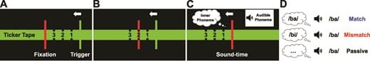
Corollary Discharge Dysfunction to Inner Speech and its Relationship to Auditory Verbal Hallucinations in Patients with Schizophrenia Spectrum Disorders
Auditory-verbal hallucinations (AVH)—the experience of hearing voices in the absence of auditory stimulation—are a cardinal psychotic feature of schizophrenia-spectrum disorders. It has long been suggested that some AVH may reflect the misperception of inner speech as external voices due to a failure of corollary-discharge-related mechanisms. We aimed to test this hypothesis with an electrophysiological marker of inner speech.
Study Design.
Participants produced an inner syllable at a precisely specified time, when an audible syllable was concurrently presented. The inner syllable either matched or mismatched the content of the audible syllable. In the passive condition, participants did not produce an inner syllable. We compared the amplitude of the N1, P2, and P3-components of the auditory-evoked potential between: schizophrenia-spectrum patients with current AVH (SZAVH+, n = 55), schizophrenia-spectrum patients without current AVH (SZAVH−, n = 44), healthy controls (HC, n = 43).

Marijuana Use Surpasses Cigarette Smoking
Rising cannabis use and falling smoking rates suggest legalization drives substitution of cannabis for cigarettes.
How does cannabis use influence cigarette smoking? This is what a recent study published in Addictive Behaviors hopes to address as a team of researchers investigated how recreational cannabis legalization has caused shifts in social dynamics, specifically regarding cigarette use. This study has the potential to help researcher better understand the social impacts of recreational cannabis legalization and the steps that can be taken to mitigate the negative impacts.
For the study, the researchers analyzed data obtained from the National Survey on Drug Use and Health for 30-day trends regarding cannabis-only use, cigarette-only use, and co-use for individuals 18 years and older and from time periods of 2015–2019, 2020, and 2021–2023. The goal of the study was to draw a connection between cannabis legalization and cigarette use, or co-use. In the end, the researchers found increases in cannabis-only use in 2015–2019, 2020, and 2021–2023 at 3.9% to 6.5%, 7.1%, and 7.9% to 10.6%, respectively. In contrast, cigarette-only use decreased during these same time periods at 15% to 12%, 10.3%, and 10.8% to 8.8%, respectively. Finally, the researchers observed consistent co-use during all three periods.
“The rising cannabis-only use across groups parallels the expanding state-level recreational cannabis legalization, increasing accessibility and normalization,” the study notes. “Conversely, continued declines in cigarette-only use align with decades of tobacco control efforts and evolving norms surrounding smoking. The relatively stable co-use trends may reflect substitution dynamics whereby some individuals replace cigarettes with cannabis, preventing co-use from rising in tandem with cannabis-only use.”