Dopamine is often called the brain’s “motivation molecule,” but for me, it represents something deeper, a window into how fragile our neurons can be. The cells that produce dopamine, known as dopaminergic neurons, are among the first to die in Parkinson’s disease, leading to the motor symptoms that gradually rob patients of movement and independence.
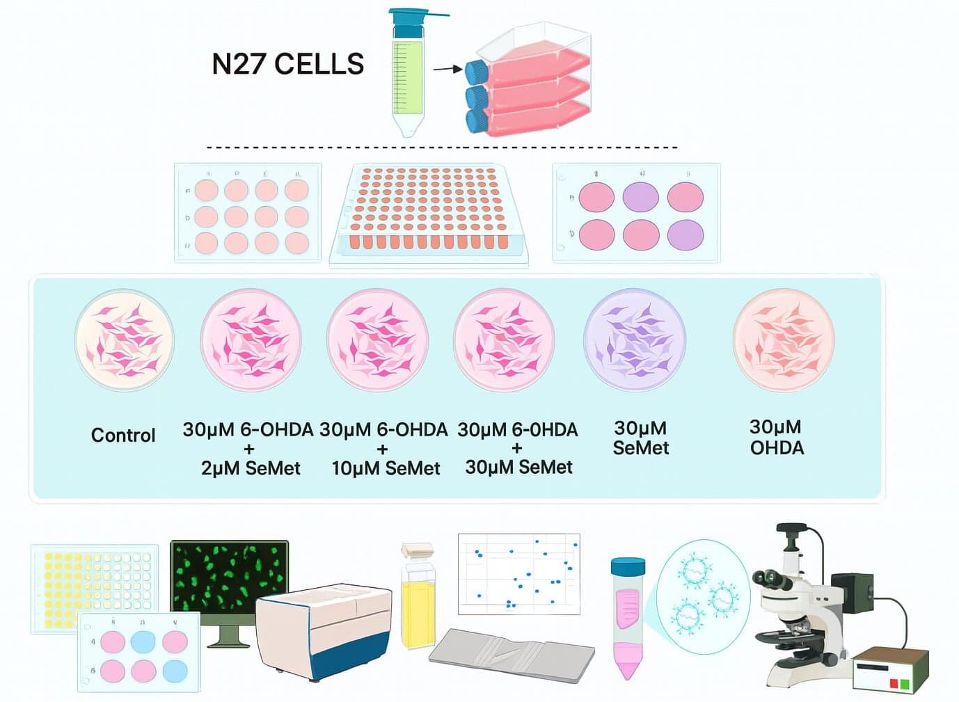
To understand what makes these neurons so vulnerable, I used an in-vitro model where I exposed N27 dopaminergic cells to 6-hydroxydopamine (6-OHDA), a toxin that triggers oxidative stress, like what occurs in the Parkinsonian brain. Then, I introduced Selenomethionine (SeMet), an organic form of selenium, to test whether this compound could counteract the damage and help the neurons survive.
Selenium has long intrigued scientists for its paradoxical nature. It is a trace element essential for antioxidant defense, yet in excess it can become toxic. I wanted to see whether a specific range of SeMet concentrations could offer meaningful protection without tipping that balance. My study, carried out at Charles University and the National Institute of Mental Health (NUDZ) in the Czech Republic, set out to define that “safe and effective window.” It is published in the journal In vitro models.