Toggle light / dark theme

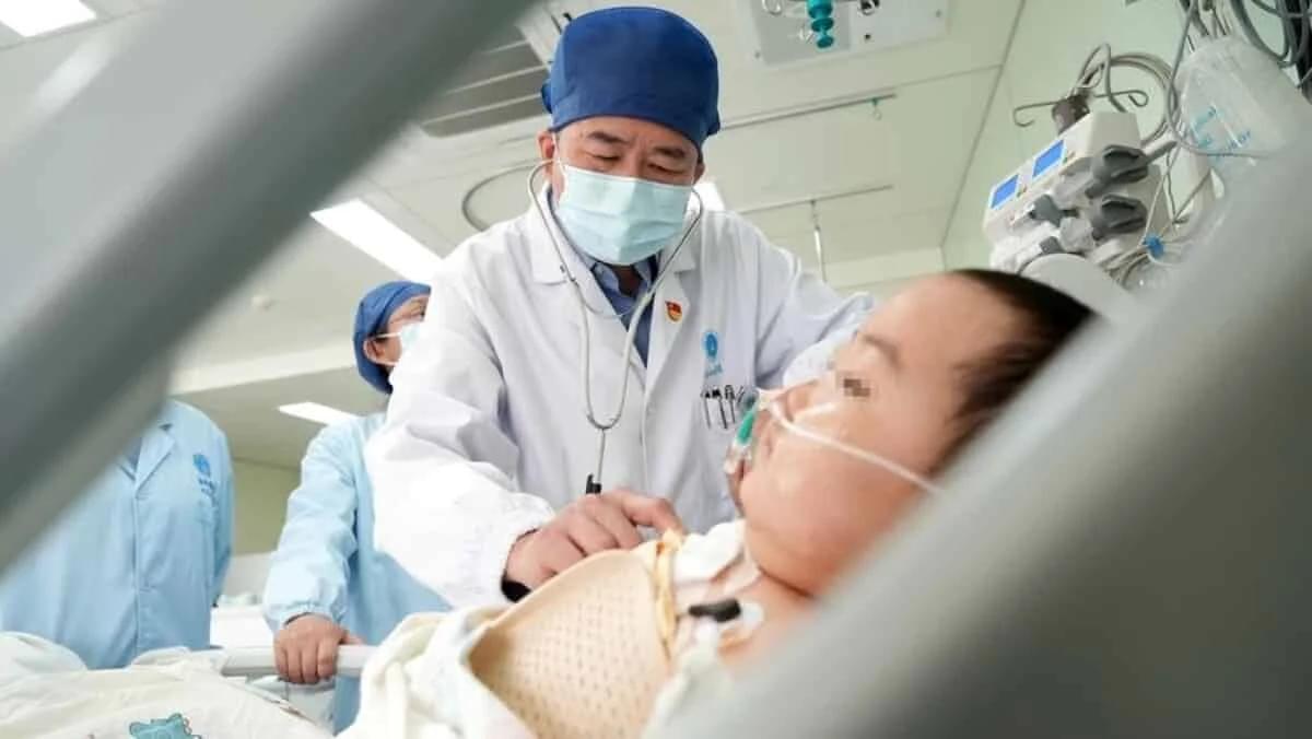
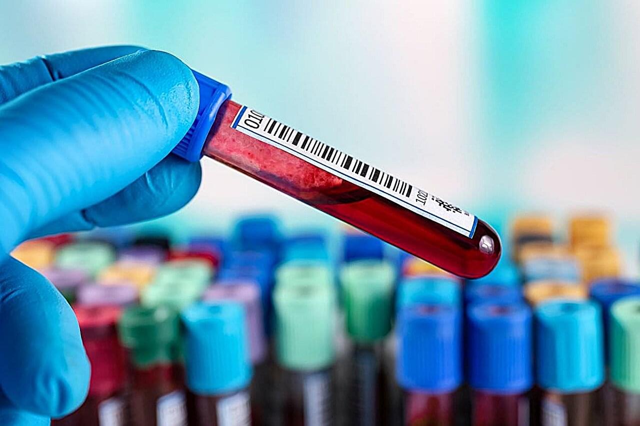
ABC News anchor Robin Roberts was brought to tears on Thursday while reporting on a new cancer treatment breakthrough. The story was about a new means of helping cancer patients find donors for blood stem cell transplants – something that Roberts herself had to deal with during her own cancer battle and subsequent health issues. She shed tears thinking of families who will not have to worry as much as she did.

Roberts presented this story along with Good Morning America co-hosts George Stephanopoulos and Lara Spencer. They explained how patients previously had to find a near perfect match for a blood stem cell transplant. This was a particular issue for people of color, but this new breakthrough more than doubles their chances of finding a match. They illustrated the effects with the real-life story of a girl who recently survived a cancer diagnosis that would have been much more dangerous beforehand. Roberts explained how broadening the list of potential donors can be so helpful – and can improve the mindsets of patients and their families as well.

Researchers from the School of Biomedical Engineering & Imaging Sciences have published a new study exploring the use of positron emission particle tracking (PEPT) in a living subject for the first time.

PEPT technology allows for the 3D localization and tracking of a single radioactive particle within large, dense, and/or optically opaque systems, which is difficult to study using other methodologies. The technology is currently used to study flows within complex mechanical systems such as large engines, industrial mixers, etc., but has not yet been translated for use in .

PEPT has previously been an unexplored area in biomedical imaging due to the lack of methods to isolate and radiolabel a single particle of a small enough size and with enough radioactivity which to would enable it to be injected and detected in a living subject.

Talk to any structural biologist, and they’ll tell you how a cool new method is taking over their field. By flash freezing proteins and bombarding them with electrons, cryo–electron microscopy (cryo-EM) can map protein shapes with near-atomic resolution, offering clues to their function and revealing bumps and valleys that drug developers can target. The technique can catch wriggly proteins in multiple configurations, and it can even capture those that have been off-limits to traditional x-ray analysis because they stubbornly resist being crystallized. Many researchers expect cryo-EM will surpass x-ray crystallography in the number of new protein structures solved next year.

Yet for all its charms, cryo-EM has flaws: The freezing process is finicky, and the microscopes are expensive. High-end machines can cost more than $5 million to buy, about as much to install, and hundreds of thousands per year to operate and maintain. Many U.S. states—and countries—don’t have a single cryo-EM microscope. “The haves and have-nots is what it is right now,” says Rakhi Rajan, a structural biologist at the University of Oklahoma, which currently lacks one.

Researchers at the Medical Research Council’s Laboratory of Molecular Biology (LMB) have been working to democratize the field. Today, in the, the U.K. team describes cobbling together a prototype cryo-EM microscope that has solved its first structures. The machine—what LMB physicist Chris Russo calls a “cheap little hatchback” rather than a “Ferrari”—could rival high-end machines in capabilities for one-tenth of the cost.

When neurons are activated in the hippocampus, not all are going to be firing at once.


Think of a time when you had two different but similar experiences in a short period. Maybe you attended two holiday parties in the same week or gave two presentations at work. Shortly afterward, you may find yourself confusing the two, but as time goes on that confusion recedes and you are better able to differentiate between these different experiences.

New research published in Nature Neuroscience reveals that this process occurs on a , findings that are critical to the understanding and treatment of memory disorders, such as Alzheimer’s disease.

Dynamic engrams store memories

In recent years, research has begun to reveal that the lines of communication between the body’s organs are key regulators of aging. When these lines are open, the body’s organs and systems work well together. But with age, communication lines deteriorate, and organs don’t get the molecular and electrical messages they need to function properly.

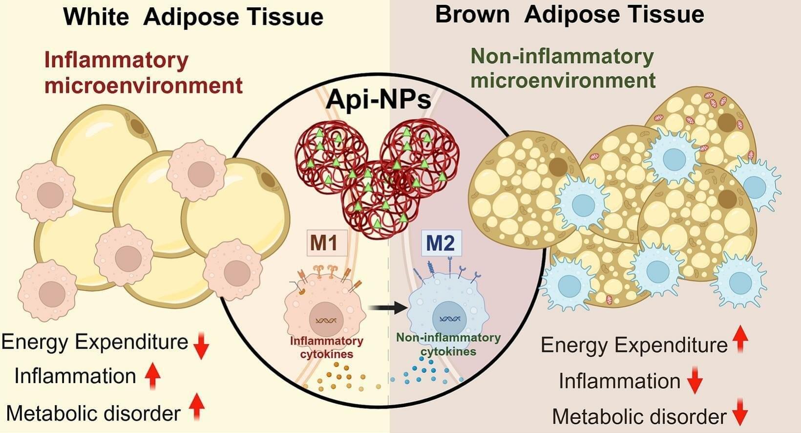
A new study from Washington University School of Medicine in St. Louis identifies, in mice, a critical communication pathway connecting the brain and the body’s fat tissue in a feedback loop that appears central to energy production throughout the body. The research suggests that the gradual deterioration of this feedback loop contributes to the increasing health problems that are typical of natural aging.

The study—published in the journal Cell Metabolism—has implications for developing future interventions that could maintain the feedback loop longer and slow the effects of advancing age.

“In recent years, the clinical development of liquid biopsies for cancer, a revolutionary screening tool, has created great optimism,” write Liz Kwo and Jenna Aronson in the American Journal of Managed Care.

At present, liquid biopsies can detect more than 50 different types of cancer. A standard visit to the doctor may eventually be able to detect cancers years before they become lethal.

In the future, even the toilet in your bathroom may be sensitive enough to detect the signs of cancer cells, enzymes and genes circulating in your bodily fluids, so that cancer becomes no more lethal than the common cold. Every time you go to the bathroom, you might be tested for cancer. The “smart toilet” might become our first line of defense.

Two insect-like robots, a mini-bug and a water strider, developed at Washington State University, are the smallest, lightest and fastest fully functional micro-robots ever known to be created.

Such miniature robots could someday be used for work in areas such as artificial pollination, search and rescue, , micro-fabrication or robotic-assisted surgery. Reporting on their work in the proceedings of the IEEE Robotics and Automation Society’s International Conference on Intelligent Robots and Systems, the mini-bug weighs in at eight milligrams while the weighs 55 milligrams. Both can move at about six millimeters a second.

“That is fast compared to other micro-robots at this scale, although it still lags behind their biological relatives,” said Conor Trygstad, a Ph.D. student in the School of Mechanical and Materials Engineering and lead author on the work. An ant typically weighs up to five milligrams and can move at almost a meter per second.

Jeff Desjardins, Editor-in-Chief of Visual Capitalist, joins OPTO Sessions to discuss the profound and far-reaching potential of CRISPR and gene editing technology, which he believes could impact fields as diverse as oncology, agriculture and materials science.

On 8 December, the US Food and Drug Administration (FDA) approved two cell-based gene therapies for the treatment of sickle cell disease. The decision marked a watershed moment in the history of healthcare, being the first time that gene therapies have won FDA approval.

One of the treatments, Casgevy, is the result of a collaboration between CRISPR Therapeutics [CRSP] and Vertex Pharmaceuticals [VRTX]. The other, Lyfgenia, was developed by bluebird bio [BLUE].