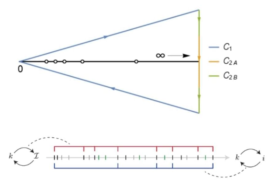
Berkeley researchers have developed a proven mathematical framework for the compression of large reversible Markov chains—probabilistic models used to describe how systems change over time, such as proteins folding for drug discovery, molecular reactions for materials science, or AI algorithms making decisions—while preserving their output probabilities (likelihoods of events) and spectral properties (key dynamical patterns that govern the system’s long-term behavior).
While describing the dynamics of ubiquitous physical systems, Markov chains also allow for rich theoretical and computational investigation. By exploiting the special mathematical structure behind these dynamics, the researchers’ new theory delivers models that are quicker to compute, equally accurate, and easier to interpret, enabling scientists to efficiently explore and understand complex systems. This advance sets a new benchmark for efficient simulation, opening the door to scientific explorations once thought computationally out of reach.
The last few weeks in longevity science have been absolutely unreal. In this episode of Longevity Science News, Emmett Short breaks down 5 bombshell breakthroughs that could reshape the future of human health in 2026 — including an FDA-approved trial aiming to reverse cellular aging, cancer vaccines eliminating brain tumors in days, the regeneration of human teeth, one-shot GLP-1 Ozempic-style gene therapies, and a shocking new discovery linking gut bacteria to multiple sclerosis.
These aren’t sci-fi predictions — these are real developments happening right now in clinical trials, biotech labs, and cutting-edge medical research. If you care about anti-aging, regenerative medicine, epigenetic reprogramming, cancer immunotherapy, GLP-1 weight loss drugs, or the future of human lifespan, this is the episode you don’t want to miss.
Hume Band 20% off with Code LSN20 https://humehealth.com/pages/hume-ban… Huma Band Review: • Best Fitness Tracker For Longevity: Hume B… JOIN LSN Patreon for exclusive access to news, tips and a community of like minded longevity enthusiasts: https://www.patreon.com/user?u=29506604 ✅ Chapters 00:00 – The Longevity Science Explosion 00:48 Hume Band 20% Off 01:02 – Exclusive Interviews 01:43 Bombshell #1: FDA Approves Age Reversal Trial (Yamanaka Factors) 04:40 – Bombshell #2: Cancer’s Worst Month Ever (Vaccines + Immunotherapy) 09:19 – Bombshell #3: The Regeneration Revolution (Cartilage, Teeth, Liver) 11:30 – Bombshell #4: The One-Shot Ozempic Gene Therapy 12:25 – Bombshell #5: Gut Bacteria Linked to Multiple Sclerosis 13:55 – Final Recap + What Breakthrough Comes Next? Links in Script David Sinclair FDA Trial Tweet https://twitter.com/davidasinclair/status/2 … FDA Greenlights Age Reset Trial (Endpoints) https://endpoints.news/exclusive-fda–… Life Biosciences Epigenetic Reprogramming Video • Reprogramming Human Life — Michael Ringel… mRNA Brain Cancer Vaccine Tweet
… ⚠️ Disclaimer: This video is for educational and informational purposes only and does not constitute medical advice. Consult a qualified clinician before making health or treatment decisions. 🔗 EXCLUSIVE INTERVIEWS & BONUS CONTENT Get extended conversations, deep dives, and behind-the-scenes research ans a YouTube Member Patreon: 👉 / u29506604 YT Membership: 👉 / @longevitysciencenews PRODUCTION CREDITS ⎺⎺⎺⎺⎺⎺⎺⎺⎺⎺⎺⎺⎺⎺⎺⎺⎺⎺⎺⎺ Executive Producer – Keith Comito @Retromancers Host, Producer, Writer – Emmett Short @emmettshort
Full huma band review: • best fitness tracker for longevity: hume B…
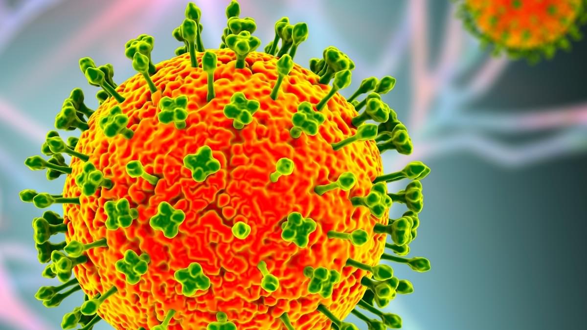
An outbreak of the deadly Nipah virus in India has put many countries in Asia on high alert, given the fatality rate in humans can be between 40% and 75%.
Several countries, including Thailand, Malaysia, and Singapore, have introduced new screening and testing measures, after at least two people died of Nipah virus in the Indian state of West Bengal this month.
But what is Nipah virus, and how concerned should we be?
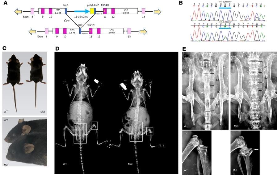
Here, Daniel H Cohn & team rescue the skeletal dysplasia phenotype of Trpv4 mutant mice—a new mouse model—using small molecule inhibition:
The figure: Reconstructed micro-CT images from WT and Co2a1-Cre/Trpv4p. R59H mutant mice showing reduction in the cervical angle (dashed red lines). The T1 vertebral body in the mutant was smaller and poorly mineralized.
2Actio Biosciences, San Diego, California, USA.
3Department of Orthopaedic Surgery, UCLA, Los Angeles, California, USA.
4Department of Nutritional Sciences, Dell Pediatric Research Institute, The University of Texas at Austin, Dell Medical School, Austin, Texas, USA.
Screening at an earlier age can help identify risk factors sooner, enabling preventive strategies that reduce long-term risk.
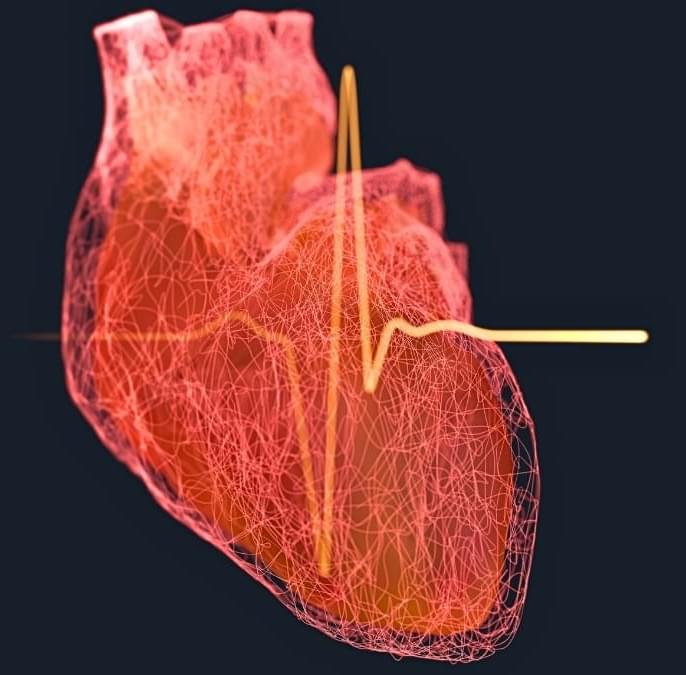
Screening for heart attack risk should be happening earlier for men, according to a new study that found the risk of cardiovascular disease starts climbing when men are in their mid-30s – significantly earlier than a similar trend is seen in women.
The US-based researchers behind the study followed the health of 5,112 people for an average of around 34 years. As the participants were healthy and aged 18–30 when the study started in the mid-1980s, the researchers could chart cases of cardiovascular disease (including strokes and heart failure) over time.
According to the data, 35 is the critical age when disparities between male and female cardiovascular disease risk start to appear. Most of the difference is driven by coronary heart disease (CHD), the most common cause of heart attacks, where fatty deposits clog up arteries, blocking blood flow.
In a recent study, researchers at the Texas A&M University Health Science Center (Texas A&M Health) identify a novel RNA molecule that plays a crucial role in preserving the integrity of a key cellular structure, the nucleolus. Their findings also suggest this molecule may influence patient survival in certain blood cancers. The work is published in the Proceedings of the National Academy of Sciences.
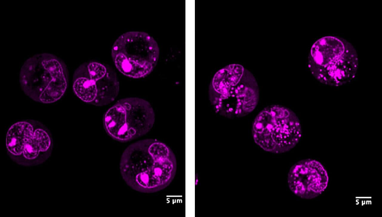
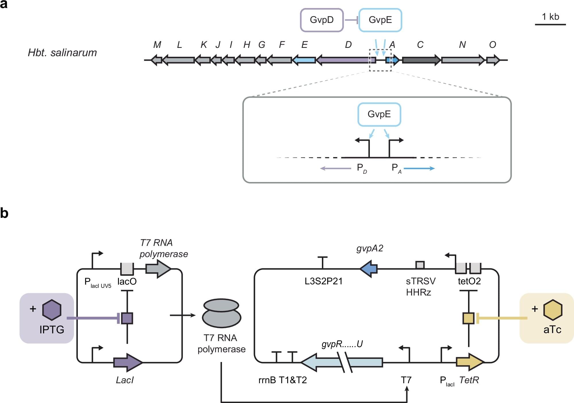
Gas vesicles are among the largest known protein nanostructures produced and assembled inside microbial cells. These hollow, air-filled cylindrical nanostructures found in certain aquatic microbes have drawn increasing interest from scientists due to their potential for practical applications, including as part of novel diagnostic and therapeutic tools. However, producing gas vesicles is a difficult task for cells in the lab, hindering the development of applications.
In a study recently published in Nature Communications, a team of researchers led by Rice University bioengineer George Lu reports the development of a new genetic regulatory system to improve cell viability during the production of gas vesicles.
“In the past few years, studies have shown that gas vesicles’ ability to reflect sound makes them useful as unique and versatile acoustic reporter systems for biomedical research and clinical applications,” said Lu, an assistant professor in the Department of Bioengineering at Rice’s George R. Brown School of Engineering and Computing.
An interdisciplinary multi-center research team led by the LKS Faculty of Medicine (HKUMed) and Faculty of Dentistry at the University of Hong Kong has constructed the world’s largest multi-omics atlas of brain metastases. This comprehensive analysis included 1,032 brain metastasis samples from diverse primary tumors, together with 82 matched primary tumors and 20 glioblastomas (a highly malignant type of brain tumor) as controls.
The findings provide a novel framework for classifying brain metastases and establish a foundation for the development of personalized treatment strategies, advancing the field of precision oncology. This research was published in the journal Nature Communications.
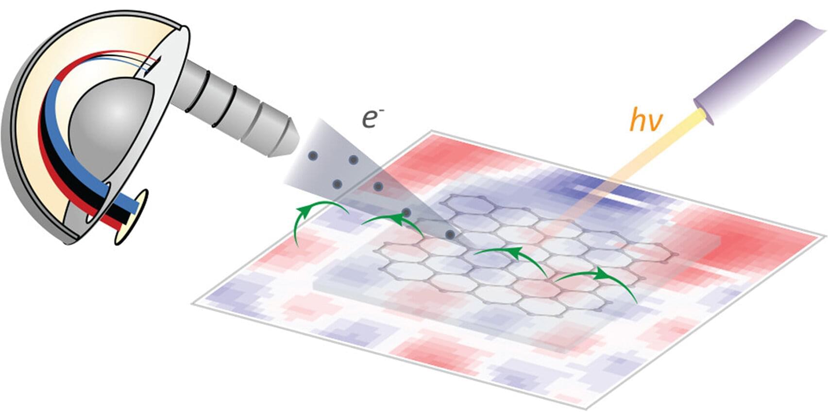
With an advanced technology known as angle-resolved photoemission spectroscopy (ARPES), scientists are able to map out a material’s electron energy-momentum relationship, which encodes the material’s electrical, optical, magnetic and thermal properties like an electronic DNA. But the technology has its limitations; it doesn’t work well under a magnetic field. This is a major drawback for scientists who want to study materials that are deployed under or even actuated by magnetic fields.
Inspired by refrigerator magnets, a team of Yale researchers may have found a solution. Their study was featured recently on the cover of The Journal of Physical Chemistry Letters.
Quantum materials —such as unconventional superconductors or topological materials—are considered critical to advancing quantum computing, high-efficiency electronics, nuclear fusion, and other fields. But many of them need to be used in the presence of a magnetic field, or even only become activated by magnetic fields. Being able to directly study the electronic structure of these materials in magnetic fields would be a huge help in better understanding how they work.
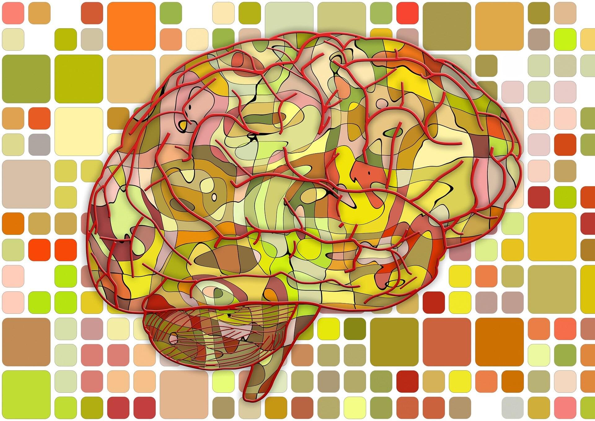
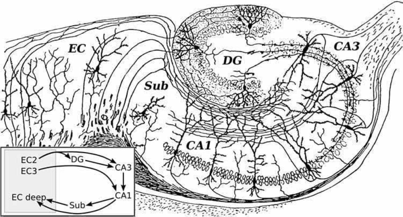
A new study finds the hippocampus reorganizes memories to predict rewards. This discovery explains how the brain learns and why Alzheimer’s affects decision-making.