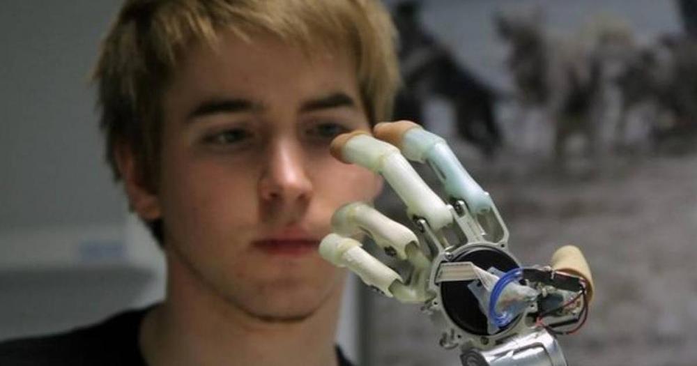
Signals from the electrical cacophony within groups of neurons inside the motor cortex were passed on to a computer running custom software for decoding.


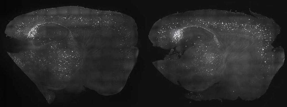
Of all the conditions that affect the elderly, one of the hardest for family and medical providers to deal with is Alzheimer’s disease. This condition impairs memory to the point that some afflicted with the condition can’t remember their loved ones. MIT researchers have found a new potential treatment that has shown promise in testing.

It is often not the lack of a cure, but the lack of will. Many great strides have been made with diabetes, when people stopped trying to develop medicines, and instead focused on how to enable the body to produce insulin.
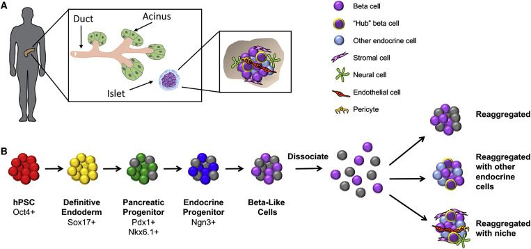
Restoration of insulin independence and normoglycemia has been the overarching goal in diabetes research and therapy. While whole-organ and islet transplantation have become gold-standard procedures in achieving glucose control in diabetic patients, the profound lack of suitable donor tissues severely hampers the broad application of these therapies. Here, we describe current efforts aimed at generating a sustainable source of functional human stem cell-derived insulin-producing islet cells for cell transplantation and present state-of-the-art efforts to protect such cells via immune modulation and encapsulation strategies.

Imagine a future where an online dating app doesn’t just match you to potential partners who meet your preferences for age, height and fondness for pinot noir, but to those with whom you’re genetically compatible. Not so much people you’re likely to have physical chemistry with – apps that make dubious claims to do that on the basis of a cheek swab already exist – but those with whom you won’t pass on a devastating genetic disease to your children.
It’s not sexy stuff; certainly not first-date conversation. Most people only discover that they’re among the four per cent who carry the same recessive genetic mutation for a rare condition, such as cystic fibrosis or Tay-Sachs, as their partner when their baby is born with it – or dies from it.
True, couples could find out their genes don’t mix after they’ve decided to have a baby and before they start trying – but how heartbreaking would that be, once they’re already in love? Far simpler never to meet in the first place, and simply to pick from the other 96 per cent with whom they can mate with abandon.

I wonder, if you can turn a 65 year old into a 40 year old, could you not turn around and give that 40 year old another treatment so they rejuvenate to an even younger state?
It is an interesting question because I might be getting near there myself! Anyway, let’s assume we develop sufficiently robust rejuvenation therapies within 20 years that they can effectively reset the clock by say 25 years with the result that a person who is chronologically 65 could be restored to a point where biologically they are 40 it begs the question could an 80 year old be effectively restored to the physiology of a 55 year old? My feeling is that the first generation treatments will in all probability be quite aggressive and invasive involving stem cell therapies, gene therapies and possibly surgical interventions to replace organs created through tissue engineering. So my concern is that whilst a 65 year old or even a fit 70 year old might easily withstand the rigours of these interventions I can’t see this applying to the average person in their 80s, whilst we are not yet at the stage where we have developed all the comprehensive therapies needed we should nevertheless keep in mind we are close and some are already approaching implementation see Suicide of aging cells prolongs life span in mice and also the video below from a few weeks back and it’s clear we are moving fast and might only be 10 to 15 years out if we keep up the current pace.
My concern is that with the senescent cell clearance a large percentage of cells in an 80 year old will be senescent so complete removal were it possible could kill them.
My thought is we might need to consider whether a less comprehensive and aggressive SENS therapy which perhaps for the sake of argument we could call MiniSENS might be useful to pull octogenarians back from the edge, perhaps by say by 10 years at which point they might be strong enough after a period of time to recover that they could handle a more intensive treatment which would yield further long term health benefits. It is just a thought but it might be something we need to think about because it would extend the age range of the people who could benefit from SENS strategies.


In some countries, progress has not always been smooth. Disease, epidemics and unexpected events are a reminder that ever-longer lives are not a given.
Meanwhile, the deaths that may preoccupy us — from terrorism, war and natural disasters — make up less than 0.5% of all deaths combined.
It will happen to all of us, but how and when we die speaks volumes about who we are and where we live.

Humans have long envied animals that are able to regenerate parts of their bodies. Arms, legs, tails, even whole chunks of the organism. Yet despite all the technology and best efforts, humans don’t have this ability. However, this could all change. Harvard University uncovered the DNA switch that controls genes for whole body regeneration. This means that one day, humans may be able to grow back lost limbs!
Many people know that certain animals are able to achieve extraordinary feats of repair, such as salamanders which grow back legs, or geckos which can shed their tails to escape predators and then form new ones in just two months. It doesn’t stop there either. Planarian worms, jellyfish, and sea anemones take this regeneration to a whole new level and can actually regenerate their entire bodies after being cut in half.
