Toggle light / dark theme

A new “toolkit” to repair damaged DNA that can lead to aging, cancer and motor neuron disease (MND) has been discovered by scientists at the Universities of Sheffield and Oxford.

Published in Nature Communications, the research shows that a protein called TEX264, together with other enzymes, is able to recognize and “eat” toxic proteins that can stick to DNA and cause it to become damaged. An accumulation of broken, damaged DNA can cause cellular aging, cancer and neurological diseases such as MND.

Until now, ways of repairing this sort of DNA damage have been poorly understood, but scientists hope to exploit this novel repair toolkit of proteins to protect us from aging, cancer and neurological disease.

Junk DNA could unlock new treatments for neurological disorders as scientists discover its breaks and repairs affect our protection against neurological disease.

The research from the University of Sheffield’s Neuroscience Institute and Healthy Lifespan Institute gives important new insights into so-called junk DNA and how it impacts on neurological disorders such as Motor Neuron Disease (MND) and Alzheimer’s.

Until now, the repair of junk DNA, which makes up 98% of DNA, has been largely overlooked by scientists, but the new study published in Nature found it is much more vulnerable to breaks from oxidative genomic damage than previously thought. This has vital implications on the development of neurological disorders.

We’ve all been there. Moments after leaving a party, your brain is suddenly filled with intrusive thoughts about what others were thinking. “Did they think I talked too much?” “Did my joke offend them?” “Were they having a good time?”

In a new Northwestern Medicine study, scientists sought to better understand how humans evolved to become so skilled at thinking about what’s happening in other peoples’ minds. The findings could have implications for one day treating such as anxiety and depression.

“We spend a lot of time wondering, ‘What is that person feeling, thinking? Did I say something to upset them?’” said senior author Rodrigo Braga. “The parts of the brain that allow us to do this are in regions of the human brain that have expanded recently in our evolution, and that implies that it’s a recently developed process. In essence, you’re putting yourself in someone else’s mind and making inferences about what that person is thinking when you cannot really know.”

While it’s well known that sleep enhances cognitive performance, the underlying neural mechanisms, particularly those related to nonrapid eye movement (NREM) sleep, remain largely unexplored. A new study by a team of researchers at Rice University and Houston Methodist’s Center for Neural Systems Restoration and Weill Cornell Medical College, coordinated by Rice’s Valentin Dragoi, has nonetheless uncovered a key mechanism by which sleep enhances neuronal and behavioral performance, potentially changing our fundamental understanding of how sleep boosts brainpower.

The research, published in Science, reveals how NREM sleep—the lighter sleep one experiences when taking a nap, for example—fosters brain synchronization and enhances information encoding, shedding new light on this sleep stage. The researchers replicated these effects through invasive , suggesting promising possibilities for future neuromodulation therapies in humans. The implications of this discovery potentially pave the way for innovative treatments for sleep disorders and even methods to enhance cognitive and behavioral performance.

The investigation involved an examination of the neural activity in multiple brain areas in macaques while the animals performed a visual discrimination task before and after a 30-minute period of NREM sleep. Using multielectrode arrays, the researchers recorded the activity of thousands of neurons across three brain areas: the primary and midlevel visual cortices and the dorsolateral prefrontal cortex, which are associated with visual processing and . To confirm that the macaques were in NREM sleep, researchers used polysomnography to monitor their brain and muscle activity alongside video analysis to ensure their eyes were closed and their bodies relaxed.

New research published by scientists at Kessler Foundation provides critical insights into the role of sleep in motor learning for individuals recovering from traumatic brain injury (TBI). The study sheds light on how sleep, specifically a short nap, influences brain activity associated with motor skill improvement, with implications for optimizing rehabilitation strategies.

The article, “Neural mechanisms associated with sleep-dependent enhancement of motor learning after brain injury”, was published in the Journal of Sleep Research. The study was led by Kessler Foundation researchers Anthony H. Lequerica, Ph.D., with additional authors Tien T. Tong, Ph.D., Paige Rusnock, Kai Sucich, Nancy Chiaravalloti, Ph.D., Ekaterina Dobryakova, Ph.D., and Matthew R. Ebben, Ph.D., and Patrick Chau, from Weill Cornell Medicine, New York.

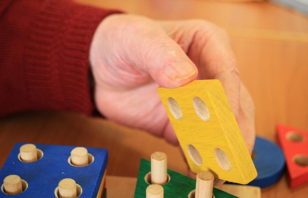
The study involved 32 individuals with TBI, randomly assigned to either a sleep or wake group following training on a motor task. The sleep group had a 45-minute nap, while the wake group remained awake, watching a documentary.

A new study by researchers at the Medical College of Wisconsin (MCW) reveals the areas of the brain where the meanings of words are retrieved from memory and processed during language comprehension. Previous neuroimaging studies had indicated that large portions of the temporal, parietal, and frontal lobes participate in processing language meaning, but it was unknown which regions encoded information about individual word meanings.

“We found that word meaning was represented in several high-level (i.e., areas that are not closely connected to primary sensory or motor areas), including the classical ‘language areas’ known as Broca’s area and Wernicke’s area,” said Dr. Leonardo Fernandino, assistant professor of neurology and biomedical engineering at MCW. “Interestingly, however, some regions not previously considered as important for language processing were among those containing the most information about word meaning.”

Additionally, they also investigated whether the neural representations of word meaning in each of these areas contained information about phenomenological experience (i.e., related to different kinds of perceptual, emotional, and action-related experiences), as several researchers had previously proposed, or whether they contained primarily information about conceptual categories (i.e., natural kinds) or about word co-occurrence statistics, as other researchers have theorized.