“Last year’s stimulus was about keeping the economy going, but these companies didn’t use these resources to retain their workers. These are companies that are polluting the environment, increasing the deadliness of the pandemic and letting go of their workers.”
Figures show 77 companies received $8.2bn under tax changes related to Covid relief and yet almost every one let workers go.
The results of this study confirm a direct link, on a molecular level, between the gut microbiome and brain function.
Summary: Consuming high levels of sugar-sweetened beverages early in life may lead to memory problems during adulthood. Researchers found, compared to rats who consumed only water, those who drank sugar-sweetened beverages had difficulties in memory recall associated with the hippocampus. The study also found a link between specific changes in gut bacteria in rats who drank sugary drinks and impaired brain function.
Source: USC
New research shows how drinking sugary beverages early in life may lead to impaired memory in adulthood.
The study, published today in Translational Psychiatry, also is the first to show how a specific change to the gut microbiome — the bacteria and other microorganisms growing in the stomach and intestines — can alter the function of a particular region of the brain.
Bipolar disorder affects millions of Americans, causing dramatic swings in mood and, in some people, additional effects such as memory problems.
While bipolar disorder is linked to many genes, each one making small contributions to the disease, scientists don’t know just how those genes ultimately give rise to the disorder’s effects.
However, in new research, scientists at the University of Wisconsin-Madison have found for the first time that disruptions to a particular protein called Akt can lead to the brain changes characteristic of bipolar disorder. The results offer a foundation for research into treating the often-overlooked cognitive impairments of bipolar disorder, such as memory loss, and add to a growing understanding of how the biochemistry of the brain affects health and disease.
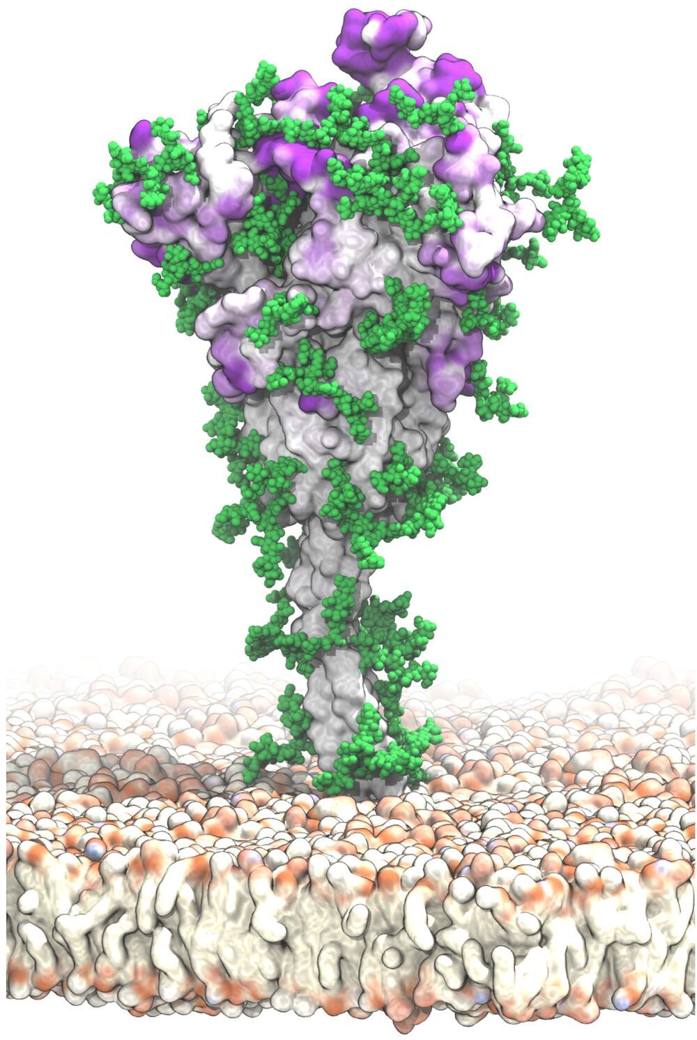
A new, detailed model of the surface of the SARS-CoV-2 spike protein reveals previously unknown vulnerabilities that could inform development of vaccines. Mateusz Sikora of the Max Planck Institute of Biophysics in Frankfurt, Germany, and colleagues present these findings in the open-access journal PLOS Computational Biology.
SARS-CoV-2 is the virus responsible for the COVID-19 pandemic. A key feature of SARS-CoV-2 is its spike protein, which extends from its surface and enables it to target and infect human cells. Extensive research has resulted in detailed static models of the spike protein, but these models do not capture the flexibility of the spike protein itself nor the movements of protective glycans—chains of sugar molecules—that coat it.
To support vaccine development, Sikora and colleagues aimed to identify novel potential target sites on the surface of the spike protein. To do so, they developed molecular dynamics simulations that capture the complete structure of the spike protein and its motions in a realistic environment.
AI plays an important role across our apps — from enabling AR effects, to helping keep bad content off our platforms and better supporting our communities through our COVID-19 Community Help hub. As AI-powered services become more present in everyday life, it’s becoming even more important to understand how AI systems may affect people around the world and how we can strive to ensure the best possible outcomes for everyone.
Several years ago, we created an interdisciplinary Responsible AI (RAI) team to help advance the emerging field of Responsible AI and spread the impact of such work throughout Facebook. The Fairness team is part of RAI, and works with product teams across the company to foster informed, context-specific decisions about how to measure and define fairness in AI-powered products.
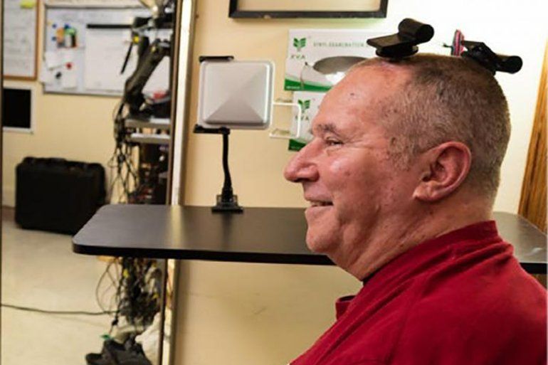
Summary: The BrainGate brain-machine interface is able to transmit signals from a single neuron resolution with full broadband fidelity without physically tethering the user to a decoding system.
Source: Brown University.
Brain-computer interfaces (BCIs) are an emerging assistive technology, enabling people with paralysis to type on computer screens or manipulate robotic prostheses just by thinking about moving their own bodies. For years, investigational BCIs used in clinical trials have required cables to connect the sensing array in the brain to computers that decode the signals and use them to drive external devices.
Gene editing has shown great promise as a non-heritable way to treat a wide range of conditions, including many genetic diseases and more recently, even COVID-19. But could a version of the CRISPR gene-editing tool also help deliver long-lasting pain relief without the risk of addiction associated with prescription opioid drugs?
In work recently published in the journal Science Translational Medicine, researchers demonstrated in mice that a modified version of the CRISPR system can be used to “turn off” a gene in critical neurons to block the transmission of pain signals [1]. While much more study is needed and the approach is still far from being tested in people, the findings suggest that this new CRISPR-based strategy could form the basis for a whole new way to manage chronic pain.
This novel approach to treating chronic pain occurred to Ana Moreno, the study’s first author, when she was a Ph.D. student in the NIH-supported lab of Prashant Mali, University of California, San Diego. Mali had been studying a wide range of novel gene-and cell-based therapeutics. While reading up on both, Moreno landed on a paper about a mutation in a gene that encodes a pain-enhancing protein in spinal neurons called NaV1.7.
Dr. Shawna Pandya MD, is a scientist-astronaut candidate with Project PoSSUM, physician, aquanaut, speaker, martial artist, advanced diver, skydiver, and pilot-in-training.
Dr. Pandya is also the VP of Immersive Medicine with the virtual reality healthcare company, Luxsonic Technologies, Director of the International Institute of Astronautical Sciences (IIAS)/PoSSUM Space Medicine Group, Chief Instructor of the IIAS/PoSSUM Operational Space Medicine course, Director of Medical Research at Orbital Assembly Construction (a company building the world’s first rotating space station providing the first artificial gravity habitat), clinical lecturer at the University of Alberta, podcast host with the World Extreme Medicine’s WEMCast series, Primary Investigator (PI) for the Shad Canada-Blue Origin student micro-gravity competition, member of the ASCEND 2021 Guiding Coalition, Life Sciences Team Lead for the Association of Spaceflight Professionals, sesional lecturer for the “Technology and the Future of Medicine,” course at the University of Alberta, and Fellow of the Explorers’ Club.
Dr. Pandya also serves as medical advisor to several space, medical and technology companies, including Mission: Space Food, Gennesys and Aquanauta, as well as the Jasper Dark Sky Festival Advisory Committee.
Dr. Pandya holds a Bsc degree in neuroscience from University of Alberta, a MSc in Space Studies from International Space University, an MD from University of Alberta, and a certification in entrepreneurship from the Graduate Studies Program at Singularity University.
Dr. Pandya is currently completing a fellowship in Wilderness Medicine (Academy of Wilderness Medicine), was granted an Honorary Fellowship in Extreme and Wilderness Medicine by the World Extreme Medicine organization in 2021, and was one of 50 physicians selected to attend the 2021 European Space Agency Space Medicine Physician Training Course. Dr. Pandya was named one of the Women’s Executive Network’s Top 100 Most Powerful Women in Canada in 2021, and a Canadian Space Agency Space Ambassador in 2021.
Dr. Pandya was part of the first crew to test a commercial spacesuit in zero-gravity in 2015. Dr. Pandya earned her aquanaut designation during the 2019 NEPTUNE (Nautical Experiments in Physiology, Technology and Underwater Exploration) mission. She previously served as Commander during a 2020 tour at the Mars Desert Research Station. Her expeditions were captured in the Land Rover short, released with the Apollo 11: First Steps film. She previously interned at ESA’s European Astronaut Center and NASA’s Johnson Space Center.
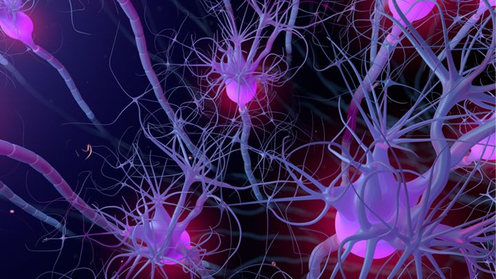
“The axons of nerve cells function a bit like a railway system, where the cargo is essential components required for the cells to survive and function. In neurodegenerative diseases, this railway system can get damaged or blocked,” Tasneem Khatib, the study’s first author, explained in a statement. “We reckoned that replacing two molecules that we know work effectively together would help to repair this transport network more effectively than delivering either one alone, and that is exactly what we found.”
Most neurodegenerative diseases are caused by multiple genetic abnormalities, making them difficult to address with gene therapy targeted at single mutations. Astellas is working on a gene therapy that expresses two proteins, and a University of Cambridge team has shown that it holds promise in glau…