Toggle light / dark theme

New MRI technology maps 20-plus brain biomarkers in a single 14-minute scan

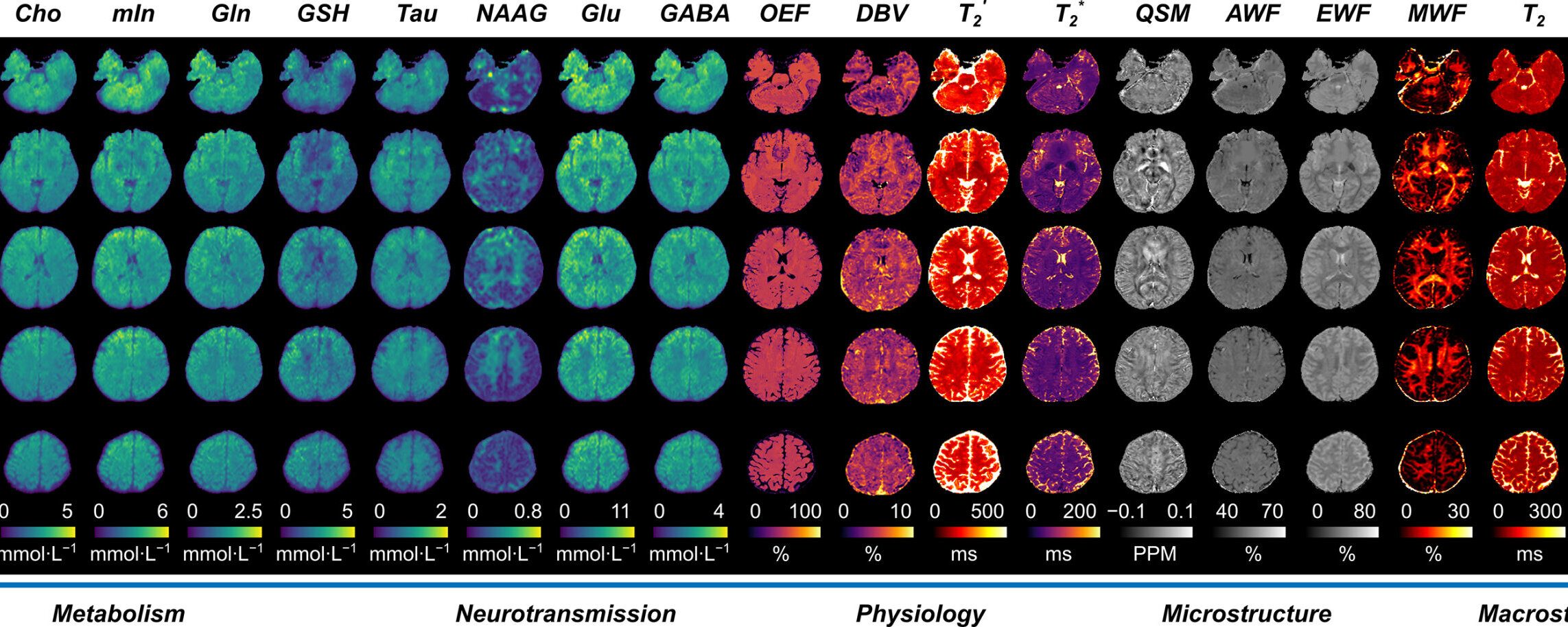
New multiplexed imaging technology using standard clinical MRI systems can simultaneously map more than 20 biomarkers in high resolution, providing a comprehensive view of the brain with a single scan.

Researchers at the University of Illinois Urbana-Champaign demonstrated the multiplexed MRI technology (MRx) by characterizing brain tumors and multiple sclerosis lesions—revealing different structural, physiological and molecular changes within the diseases. Led by Zhi-Pei Liang, a professor of electrical and computer engineering and a member of the Beckman Institute for Advanced Science and Technology at the U. of I., the team has reported its findings in the journal Nature.

“MRx can be a powerful tool for noninvasive tissue characterization, helping to advance personalized, precision and predictive medicine,” Liang said. “By providing rich, multidimensional biomarkers to capture disease progression and treatment response, this capability could open new opportunities for more precise diagnosis, individualized treatment planning and improved patient outcomes.”


Study of a Million Blood Cells Helps Explain Why Women Face More Autoimmune Disease

Autoimmune diseases, where the body’s own immune system mistakenly goes on the attack, are much more common in women – and a new study analyzing more than 1.25 million blood cells goes a long way to explaining why.

The analysis, led by a team from the Garvan Institute of Medical Research in Australia, revealed over 1,000 genetic ‘switches’ in immune cells that work differently depending on sex.

In short, these variations in gene activity mean that inflammatory pathways that respond to threats are likely to be busier in women, leading to a greater risk of conditions like lupus and multiple sclerosis.

A gene that keeps intestinal stem cells stable offers insight into how tissues repair themselves

Years before he conducted the research that would earn him a Nobel Prize in Physiology and Medicine, Shinya Yamanaka, MD, Ph.D., was a postdoctoral scientist at Gladstone Institutes, studying genes. There, he helped discover a gene (now called eIF4G2) that’s essential for early embryonic development.

Then, the story pauses. Without the technology needed to develop an animal model to further investigate the gene, Yamanaka moved on to develop induced pluripotent stem (iPS) cells—adult cells that have been reprogrammed into an embryonic state. That work earned him the Nobel Prize, but he never forgot his first gene.

Now, 30 years since his postdoc, Yamanaka has circled back to eIF4G2.

LED light unlocks 3D optical fingerprints inside materials without lasers

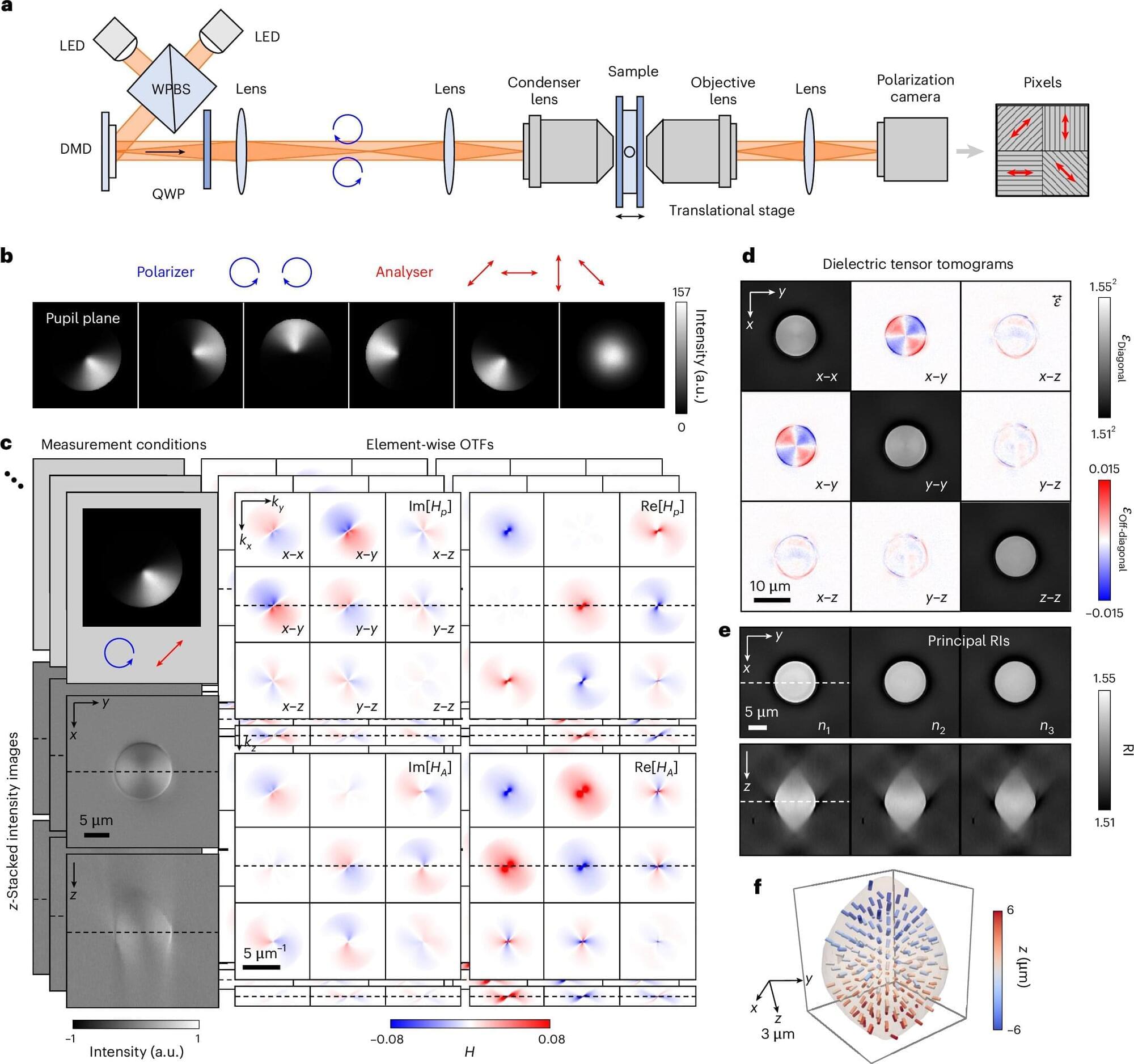
Researchers have developed, for the first time in the world, incoherent dielectric tensor tomography (iDTT), a technology that can read complex three-dimensional optical fingerprints inside materials using only everyday LED illumination.

The study is published in Nature Photonics, and the research team was led by Professor YongKeun Park of the Department of Physics, in collaboration with Professor Seung-Mo Hong’s team at Asan Medical Center and Professor Seokwoo Jeon’s team at Korea University.

Some materials possess an inherent property called optical anisotropy, in which the refractive index changes depending on the direction in which light passes through. This is a decisive optical fingerprint that reveals the internal structure and molecular arrangement of the material.

Leukemia stem cells cause treatments to fail, but findings open new avenues to overcome resistance

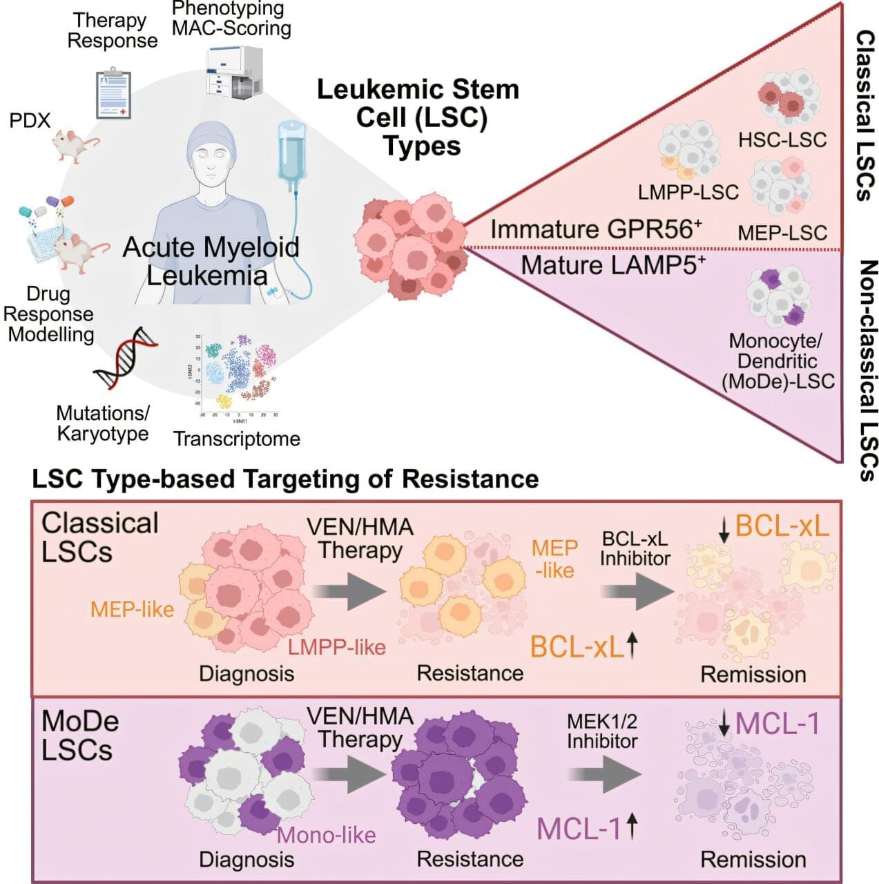
Scientists from the German Cancer Research Center (DKFZ) and the HI-STEM Stem Cell Institute have deciphered a key mechanism that contributes to treatment failure in acute myeloid leukemia (AML). They show that there are not just one, but four different subtypes of leukemia stem cells. This diversity could explain why one of the most important AML drugs does not work sufficiently in some patients or loses its effectiveness over time—resulting in the return of leukemia.

This discovery lays an important foundation for more precise and long-term successful treatment strategies that could specifically overcome resistance mechanisms. The findings are published in the journal Cell Stem Cell.

Acute myeloid leukemia (AML) is an aggressive form of blood cancer that primarily affects older people and often has a poor prognosis despite improved therapies. In recent years, the targeted drug venetoclax has significantly improved treatment. In combination with other drugs, venetoclax often shows good therapeutic success in AML and will, at least in part, replace highly aggressive chemotherapy in the future. However, AML returns in nearly all patients—usually because individual cancer stem cells become resistant to the drug.

Magnetic checkerboard separates microparticles by size and sends them along different paths

A team of researchers from the Universities of Tübingen, Bayreuth, and Kassel, and the Polish Academy of Sciences has developed a method for precisely controlling the movement of magnetic microparticles based on their size. These suspended particles, known as colloidal particles, range in size from a few tens of nanometers to several micrometers. Controlling them is important for applications such as drug delivery, medical laboratory tests, and the synthesis of new materials. The team’s study has now been published in Physical Review Letters.

The new method involves positioning microparticles above a magnetic layer that is patterned like a chessboard. In previous studies, magnetic transportation of the colloidal particles was limited to a specific height. At this distance, although the magnetic forces appear to balance each other out, the particles move regardless of their size. Therefore, it was not possible to control the particles specifically based on their size.

/* */