No matter the size or severity, wounds on human skin are difficult to monitor while they heal. Biopsies disrupt the wound site and are too invasive for routine, repeated monitoring, and most medical imaging devices that could do the job are large, expensive, and booked up with more pressing diagnostics. Clinicians typically resort to visual inspection or quick measurements of the wound’s size over time.
Based on research completed as part of a multi-year collaboration with Nokia Bell Labs, biomedical engineers at Duke University are developing a solution. Using a custom-built optical coherence tomography (OCT) imaging system together with artificial intelligence (AI) models grounded in a deep understanding of tissue regeneration, researchers have shown they can accurately and objectively measure the progress of wounds healing over time.
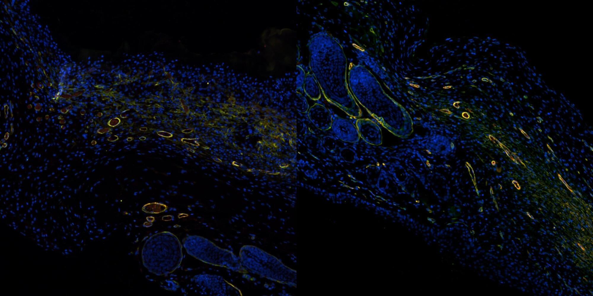
Using their new approach, the researchers also show that a hydrogel under development to improve wound healing works better with stiffer mechanical properties. The results are a two-for-one boon in a challenging area for both clinicians and researchers.