These are the molecular machines inside your body that make cell division possible. Animation by Drew Berry at the Walter and Eliza Hall Institute of Medical Research. http://wehi.tv.
Special thanks to Patreon supporters: Joshua Abenir, Tony Fadell, Donal Botkin, Jeff Straathof, Zach Mueller, Ron Neal, Nathan Hansen.
Every day in an adult human roughly 50–70 billion of your cells die. They may be damaged, stressed, or just plain old — this is normal, in fact it’s called programmed cell death.
To make up for that loss, right now, inside your body, billions of cells are dividing, creating new cells.
And cell division, also called mitosis, requires an army of tiny molecular machines. DNA is a good place to start — the double helix molecule that we always talk about.
In this video students of the Maastricht Science Program NanoBiology Course 2020, show their explanation of the SARS-CoV-2 viral budding. Using CellPAINT, UCFS Chimera and their creativity they explain the nanobiology of how the SARS-CoV-2 virion can bud and leave the cell.
Viruses are not living things. They are just complicated assemblies of molecules, in particular macromolecules such as proteins, oligonucleotides, combined with lipids and carbohydrates. A virus cannot function or reproduce by itself. It needs a host cell.
When a virus enters the host cell, a series of chemical reactions occur that lead to the production of new viruses. A virus needs to find a host cell, attach to it, enter it, and reprogram it such that it will replicate its genome and produce new proteins that allow the assembly of a new virus. Once new viruses have been assembled, they need to get out of the original host cell, on their way to the next host cell they can exhaust. Some viruses have an easy way out: they use up all the resource of the host cells until it dies and lyse. This would only work for naked viruses such as polyomavirus and adenovirus, which lacks a lipid membrane.
Washing hands has been a standard measure since the start of this COVID-19 pandemic. The soap will disintegrate the lipid envelop of the SARS-CoV2 viral particles, as this is an enveloped virus. Enveloped viruses need envelopment, a process in which the capsids become surrounded by a lipid bilayer. This process takes place prior to release. Two mechanisms for envelopment exist. First, envelopment can proceed sequentially after the completion of capsid assembly. The fully assembled capsids are recruited to the membrane by interaction of the viral capsids with viral envelope glycoprotein. Examples of this include herpesvirus and hepatitis B virus. Secondly, the envelopment can occur simultaneously with the capsid assembly. Retrovirus is the representative of this coupled mechanism.
Where does the membrane for the envelopment come from? Some viruses, such as retrovirus and influenza virus, using the plasma membrane as the site of envelopment, whereas others, such as herpesvirus and hepatitis B, use the endoplasmic reticulum (ER) and Golgi bodies as the site of envelopment.
Enveloped viruses are released from the infected cell via exocytosis, a process which is often also called budding. Viruses exploit cellular mechanisms to produce their own progeny extracellularly. For example, the budding of retroviral Gag is facilitated by ESCRT complexes, which are normally involved in the multi-vesicular bodies (MVB) pathway. How does SARS-CoV2 release its offspring from the infected cell? Can we interfere with these steps such that we attack the virus at each step of its life cycle?
Full Credits: Production: Birdo. Script: Dr. Paulo César Naoum, Aliá F. M. Naoum. Direction: Luciana Eguti, Paulo Muppet. Storyboard: Antonio Linhares, Pedro Eboli. Design and Animation: Antonio Linhares, Pedro Eboli, Rafael Gallardo. Sound design: Antonio Linhares.
“Like a lock and key” — this is the description of how viruses can get into our cells. Viruses use special proteins on their surface to enter cells. They do this because they need our cells to reproduce. But viruses can only enter certain cells. They use proteins on their surface that act like keys to unlock human cell receptors to invade and infect cells.
The Vaccine Makers Project (VMP) is the classroom-based program of the Vaccine Education Center at the Children’s Hospital of Philadelphia (VEC at CHOP). The Center’s team is composed of scientists, physicians, mothers and fathers devoted to the study and prevention of infectious diseases. The Center was launched in October 2000 to provide accurate, comprehensive and up-to-date information about vaccines and the diseases they prevent. The VMP program is committed to public education about vaccine science via scientifically supported, historically accurate, and emotionally compelling content.
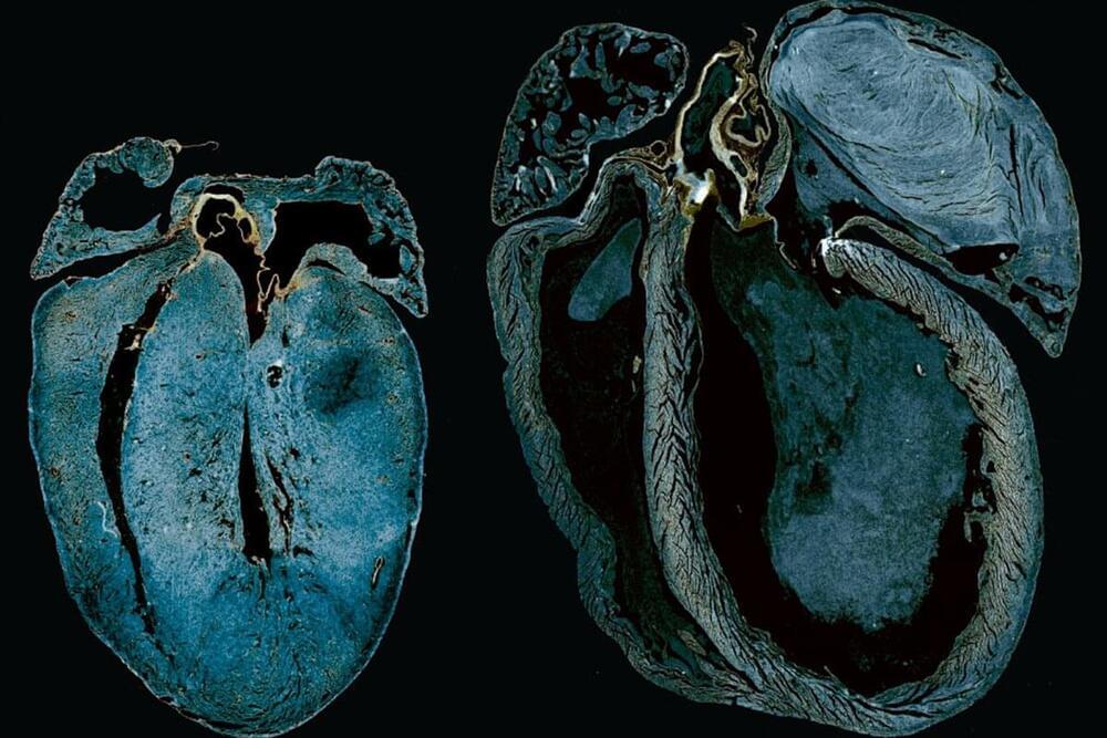
Heart failure is often identified only when the heart has already deteriorated. This is in large part because the cause is unknown for about 70% of people who experience heart failure.
Researchers at The Hospital for Sick Children (SickKids) have discovered that one of the earliest signs of heart failure is a change in how the heart produces energy, with findings offering a potential way to preempt heart failure before the heart begins to deteriorate.
Led by Dr. Paul Delgado-Olguín, a scientist in the Translational Medicine program, the research may also help to explain the diversity of causes underlying heart failure.
ChatGPT has passed the gold-standard exam required to practice medicine in the US — amid rising concerns AI could put white-collar workers out of jobs.
The artificial intelligence program scored between 52.4 and 75 percent across the three-part Medical Licensing Exam (USMLE). Each year’s passing threshold is around 60 percent.
Researchers from tech company AnsibleHealth who did the study said: ‘Reaching the passing score for this notoriously difficult expert exam, and doing so without any human reinforcement, marks a notable milestone in clinical AI maturation.’
Dr. Craig Kaplan discusses Artificial Intelligence — the past, present, and future. He explains how the history of AI, in particular the evolution of machine learning, holds the key to understanding the future of AI. Dr. Kaplan believes we are on an inexorable path towards Artificial General Intelligence (AGI) which is both an existential threat to humanity AND an unprecedented opportunity to solve climate change, povery, disease and other challenges. He explains the likely paths that will lead to AGI and what all of us can do NOW to increase the chances of a positive future.
Chapters. 0:00 Intro. 0:22 Overiew & summary. 0:45 Antecedents of AI 1:15 1956: Birth of the field / Dartmouth conference. 1:33 1956: The Logic Theorist. 1:58 1986: Backprogation algorithm. 2:26 2016: SuperIntelligent AI / Alpha Go. 2:51 Lessons from the past. 3:59 Today’s “Idiot Savant” AI 4:45 Narrow vs. General AI (AGI) 5:15 Deep Mind’s Alpha Zero. 6:19 Demis Hassabis on Alpha Fold. 6:47 Alpha Fold’s amazing performance. 8:03 OpenAI’s ChatGPT 9:16 OpenAI’s DALL-E2 9:50 The future of AI 10:00 AGI is not a tool. 10:30 AGI: Intelligent entity. 10:48 Humans will not be in control. 11:16 The alignment problem. 11:45 Alignment problem is unsolved! 12:45 Likely paths to AGI 13:00 Augmented Reality path to AGI 13:26 Metaverse / Omniverse path to AGI 14:20 AGI: Threat AND Opportunity. 15:10 Get educated — books. 15:48 Get educated — videos. 16:20 Raise awareness. 16:44 How to influence values of AGI 17:52 No guarantees, we must do what we can. 18:47 AGI will learn our values. 19:30 Wrap up / contact info.
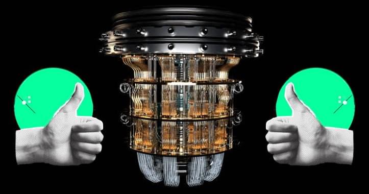
Researchers in the US developed a new energy-based benchmark for quantum advantage and used it to demonstrate noisy intermediate-scale quantum (NISQ) computers that use several orders of magnitude less energy than the world’s most powerful supercomputer. Quantum computing is a branch of computer science that focuses on the development of technologies based on quantum theory principles.
Quantum computing solves problems that are too complex for classical computing by utilizing the unique properties of quantum physics. The question of whether a quantum computer can perform calculations beyond the reach of even the most powerful conventional supercomputer is becoming increasingly relevant as quantum computers become larger and more reliable. This ability, dubbed “quantum supremacy,” marks the transition of quantum computers from scientific curiosity to useful devices. Scientists predict that Quantum computing is better than supercomputers as it performs tasks a million times faster. Quantum computers can handle complex calculations easily because they are built based on quantum principles that go beyond classical physics.
Quantum computers and supercomputers are extremely powerful machines used for complex calculations, problem solving, and data analysis. While both have the potential to revolutionize computing technology, they have significant speed and capability differences. In 2019, Google’s quantum computer performed a calculation that would take the world’s most powerful computer 10,000 years to complete. It is the seed for the world’s first fully functional quantum computer, which will be capable of producing better medicines, developing smarter artificial intelligence, and solving cosmic mysteries. Theoretical physicist John Preskill proposed a formulation of quantum supremacy, or the superiority of quantum computers, in 2012. He dubbed it the moment when quantum computers can perform tasks that ordinary computers cannot. To quickly crunch large amounts of data and achieve a single result, supercomputers employ a traditional computing approach with multiple processors.