Toggle light / dark theme

Dr Veronica Rodriguez-Bravo PhD — Nuclear Pore Complexes — Sidney Kimmel Cancer Center, Philadelphia

Nuclear Pore Complexes and Genome Integrity — Dr. Veronica Rodriguez-Bravo Ph.D., Sidney Kimmel Cancer Center — Jefferson Health, Thomas Jefferson University.


Dr. Veronica Rodriguez-Bravo, PhD, is Assistant Professor, Department of Cancer Biology, at the Sidney Kimmel Cancer Center, Thomas Jefferson University, in Philadelphia, PA, USA. (https://sidneykimmelcancercenter.jeffersonhealth.org/)

Dr. Rodriguez-Bravo obtained her PhD in Pathology and Cell Biology (Summa Cum Laude) from the University of Barcelona in 2007, where she also received the Extraordinary Doctorate Award for her studies on the distinct DNA replication checkpoint mechanisms of tumor cells. During her postdoctoral training at the Experimental Oncology Department of the University Medical Center of Utrecht (UMC, The Netherlands) and at the Molecular and Cell Biology Programs of Memorial Sloan Kettering Cancer Center (MSKCC, New York), she specialized in the study of chromosome segregation during mitosis and the role of nuclear pores in genome integrity maintenance.

Dr. Rodriguez-Bravo’s post-doctoral work allowed her to apply genome-editing techniques crucial to dissect the function of mitotic and nuclear pore proteins in chromosomal stability and resulted in the recognition with the Memorial Sloan Kettering Cancer Center Postdoctoral Research Award.

Dr. Rodriguez-Bravo’s research focuses on the study of genome integrity maintenance mechanisms and the relationship of defects in cell division to cancer pathogenesis with special emphasis in the pathways contributing to cancer cells’ more aggressive phenotypes.
Cannabinoid Pathway Linked to Psychiatric Disorders

“Cannabis may contribute to increased risk for mental disorders, which has actually been shown in schizophrenia,” Penzes said. “Conversely, cannabis could be beneficial in some brain disorders, which prompted trials of medical marijuana in patients with autism.”


Summary: Findings reveal a role the endocannabinoid system plays in a range of psychiatric disorders, including schizophrenia, bipolar disorder, and ASD.

Source: Northwestern University

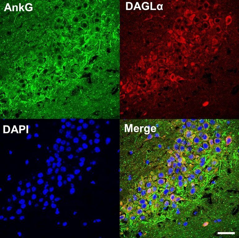
Northwestern Medicine scientists discovered an unexpected connection between a synapse protein that has been implicated in neuropsychiatric disorders and the endocannabinoid pathway, according to a study published in Biological Psychiatry.

These findings suggest a role for the endocannabinoid system in conditions including bipolar disorder, according to Peter Penzes, PhD, the Ruth and Evelyn Dunbar Professor of Psychiatry and Behavioral Sciences, professor of Physiology and Pharmacology, and senior author of the study.

A noninvasive technique for neurological conditions

Indiana University School of Medicine researchers are developing a new, noninvasive brain stimulation technique to treat neurological disorders, including pain, traumatic brain injury (TBI), epilepsy, Parkinson’s disease, Alzheimer’s disease and more.

“Given the increasing use of stimulation in human brain study and treatment of neurological diseases, this research can make a big impact on physicians and their patients,” said Xiaoming Jin, Ph.D., associate professor of anatomy, cell biology and physiology.

When someone experiences a , nerve injury, or neurodegeneration, such as in epilepsy and TBI, there is damage to the brain which can lead to loss and damage of nerve or neurons and development of hyperexcitability that underlies some neurological disorders such as neuropathic pain and epilepsy.

Harvard, MIT researchers create COVID-19 diagnosing mask

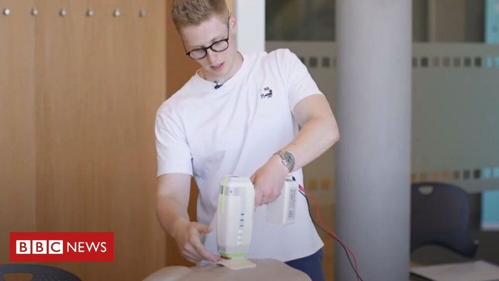
Researchers from Harvard University and the Massachusetts Institute of Technology, both based in Cambridge, Mass., have created small diagnostic biosensors that can be inserted into face masks and can diagnose COVID-19 within 90 minutes, The Mercury News reported June 29.

The insertable biosensors detect the virus from a wearer’s breath, producing easy to read results similar to those of an at-home pregnancy test. If the coronavirus is present, the system changes the pattern of lines in the readout strip.

To activate the test, the wearer pushes a button on the mask to release a small amount of water into the system, which activates the test.

Stem cell-based biological tooth repair and regeneration

Stem cells for teeth repair.


Teeth exhibit limited repair in response to damage, and dental pulp stem cells probably provide a source of cells to replace those damaged and to facilitate repair. Stem cells in other parts of the tooth, such as the periodontal ligament and growing roots, play more dynamic roles in tooth function and development. Dental stem cells can be obtained with ease, making them an attractive source of autologous stem cells for use in restoring vital pulp tissue removed because of infection, in regeneration of periodontal ligament lost in periodontal disease, and for generation of complete or partial tooth structures to form biological implants. As dental stem cells share properties with mesenchymal stem cells, there is also considerable interest in their wider potential to treat disorders involving mesenchymal (or indeed non-mesenchymal) cell derivatives, such as in Parkinson’s disease.

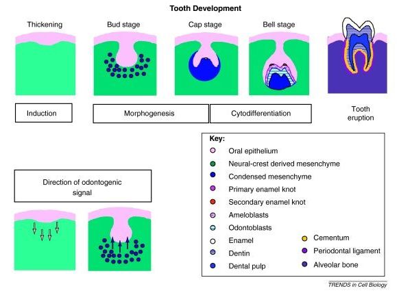
Teeth are complex organs containing two separate specialized hard tissues, dentine and enamel, which form an integrated attachment complex with bone via a specialized (periodontal) ligament. Embryologically, teeth are ectodermal organs that form from sequential reciprocal interactions between oral epithelial cells (ectoderm) and cranial neural crest derived mesenchymal cells. The epithelial cells give rise to enamel forming ameloblasts, and the mesenchymal cells form all other differentiated cells (e.g., dentine forming odontoblasts, pulp, periodontal ligament) (Box 1). Teeth continue developing postnatally; the outer covering of enamel gradually becomes harder, and root formation, which is essential for tooth function, only starts to occur as part of tooth eruption in children.

Tooth development is traditionally considered a series of stages that reflect key processes ( Figure I ). The first step is induction, in which signals from the epithelium to the mesenchyme initiate the developmental process. As localized proliferation of the dental epithelial cells takes place, the cells form a bud around which the mesenchymal cells condense. Differentiation and localized proliferation of the epithelial cells in the bud leads to the cap stage. It is at this stage that crown morphogenesis is initiated by the epithelial signalling centre, an enamel knot regulating the folding of the epithelium. By the bell stage, the precursors of the specialized tooth cells, ameloblasts, coordinate enamel deposition, and odontoblasts, which produce dentine, are formed. Tooth eruption involves the coordination of bone resorption and root development, and occurs postnatally.

Translating Blood Plasma Dilution To Humans | Drs. Irina & Mike Conboy Interrview Series Ep3

The Gist: They think they can start wider human trials soon which would last 2 years then have a product in 3 to 4 years.


In this video, Drs Irina and Mike Conboy talk how TPE, therapeutic plasma exchange is already available as an FDA approved procedure and the plans to extend the usage to include more age related diseases. We also discuss the company that they have formed IMU

Our guests today are Drs. Irina and Michael Conboy of the Department of Bioengineering at the University of California Berkeley. their discovery of the rejuvenating effects of young blood through parabiosis in a seminal paper published in Nature in 2005 paved the way for a thriving field of rejuvenation biology. The Conboy lab currently focuses on broad rejuvenation of tissue maintenance and repair, stem cell niche engineering, elucidating the mechanisms underlying muscle stem cell aging, directed organogenesis, and making CRISPR a therapeutic reality.

Papers mentioned in this video.
Plasma dilution improves cognition and attenuates neuroinflammation in old mice.
https://pubmed.ncbi.nlm.nih.gov/33191466/
Rejuvenation of three germ layers tissues by exchanging old blood plasma with saline-albumin.
https://pubmed.ncbi.nlm.nih.gov/32474458/
Rejuvenation of aged progenitor cells by exposure to a young systemic environment.
https://pubmed.ncbi.nlm.nih.gov/15716955/

************************************************************

Guardian Deletes Fake News Article on Life Extension

Did you know that life extension is impossible? I didn’t until the keen minds over at the Guardian informed me as such. Fortunately before I decided to give up my career in regenerative medicine I decided to look into exactly how the Guardian came to such a revelation. What I discovered was so earth shattering that it rocked my very understanding of the world, and made me question everything I thought I knew. That realisation was that, despite their consistent insistence to the contrary, the media lies, a lot.

Ok maybe this was not as shocking as I made it out to be, in fact it’s pretty widely known by now that the media is generally no stranger to the odd lie here and there. If you are not familiar with the story allow me to give you a recap. A recent study was conducted in order to find out if human lifespans have actually increased due to advanced in medicine. To do this, scientists used statistical models to remove non-age related causes of death from historical records (such as murder, death in child birth, plague etc) in order to determine what the uninterrupted human lifespan is, and if it has increased over time. What was found is that our medical science is yet to fundamentally extend human lifespan. This comes as no surprise to anyone in the field of longevity research as we know full well that none of our current medical treatments address causes of ageing. What this study does not conclude however is that life extension is impossible.

Molecular machines talk to living cells for the first time

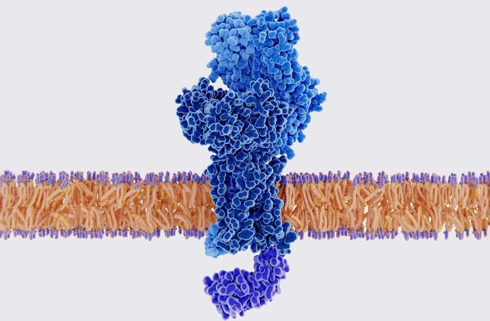
For the first time, an artificial molecular motor has been created that can ‘talk’ to living cells – by gently pulling their surface with enough physical force to elicit a biochemical response. The approach could help scientists decode the language that cells use to communicate with each other in tissues.

‘There is a mechanical language in the form of physical forces applied by the cells themselves, and we want to understand what information is communicated and what the consequences are,’ explains Aránzazu del Campo, who led the study at the Leibniz Institute for New Materials, Germany. ‘Ultimately, we want to be able to provide signals to cells and guide their function when they are not able to do that by themselves in disease cases.’

Usually, studying how cells communicate by sensing mechanical stimuli and producing biochemical responses requires prodding them with pipettes or the tip of an atomic force microscope. However, this doesn’t work at the more complex tissue level.