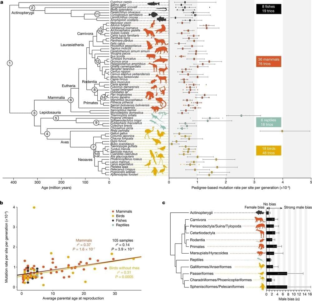
Toggle light / dark theme

A tale of GS-441524, the remdesivir sister drug that cures coronavirus (FIP) in cats, but that Gilead refused to develop for fear it would mess up the approval process of remdesivir.

Yes, it a tale of capitalism on steroids, and the FDA on drugs. It’s the kind of thing that may well kill you and your family, but you will never know about it (unless you read about it in The Atlantic, or some obscure journal).


Cat owners are resorting to China’s underground marketplace to buy antivirals for a feline coronavirus.

With more and more people contracting COVID-19, physician-scientists around the world are looking at existing drugs as potential treatments for the novel coronavirus.

Among them are cancer researchers launching new clinical trials or participating in multi-site ones to see whether drugs proven to be effective for another disease, particularly cancer, can help in treating COVID-19 cases.

At Miami Cancer Institute, part of Baptist Health South Florida, researchers are conducting several trials to investigate whether existing cancer drugs might provide some kind of therapy for COVID-19. The rationale: Cancer patients have similar immune system problems as coronavirus patients — namely, dangerous inflammation — and these medications effectively attack that problem.

Oxford University scientists leading the global search for a coronavirus vaccine are to recruit “very healthy” over-55s to help with clinical trials.

The next phase of testing will focus on how older adults’ immune systems respond, the Oxford vaccine group said on Friday.

Scientists are looking for 10,260 people from across the UK to take the jab, considered a front-runner in the world race for a vaccine.

Summary: Tau spreads through the human brain via neural communication pathways. The spread is accelerated by the presence of amyloid-beta.

Source: Lund University

Toxic versions of the protein tau are believed to cause death of neurons of the brain in Alzheimer’s disease. A new study published in Nature Communications shows that the spread of toxic tau in the human brain in elderly individuals may occur via connected neurons. The researchers could see that beta-amyloid facilitates the spread of toxic tau.

Circa 2007


This chapter describes detection of explosives by terahertz Imaging ™. There has been an amplified interest in terahertz (THz) detection for imaging of covered weapons, explosives, chemical and biological agents. THz radiation is readily transmitted through most nonmetallic and nonpolar mediums. This process enables the THz systems to see through concealing barriers, which includes packaging, corrugated cardboard, clothing, shoes, book bags, and such others to find potentially dangerous materials concealed within. Apart from many materials of interest for security applications, which include explosives, chemical agents, and other such biological agents that have characteristic THz spectra which can be used for fingerprint testing and identify concealed materials. The Terahertz radiation poses either no or minimal health risk to either a suspect being scanned by a THz system or the system’s operator. As plastic explosives, fertilizer bombs, and chemical and biological agents increasingly become weapons of war and terrorism, and the trafficking of illegal drugs increasingly develops as a systemic threat, effective means for rapid detection, and an identification of these threats are required. One proposed solution for locating, detecting, and characterizing concealed threats is to use THz electromagnetic waves to spectroscopically detect and identify concealed materials through their characteristic transmission or reflectivity spectra in the range of 0.5–10 THz.

Now abandoned bikes are strewn across the streets, the leather-covered massage chairs are empty amid worries over cleanliness and people are ordering more of their daily necessities online and avoiding malls altogether. After weeks of lockdowns and social distancing measures to combat the spread of the virus, many people are asking whether this fabled part of China’s shiny new tech-driven economy will ever recover its former glory.


Experts say that consumer behaviour has changed irrevocably as a result of Covid-19 – and that the sharing economy must adapt.

The most common organism in the oceans, and possibly on the entire planet, is a family of single-celled marine bacteria called SAR11. These drifting organisms look like tiny jelly beans and have evolved to outcompete other bacteria for scarce resources in the oceans.

We now know that this group of thrives despite—or perhaps because of—the ability to host viruses in their DNA. A study published in May in Nature Microbiology could lead to new understanding of viral survival strategies.

University of Washington oceanographers discovered that the that dominate seawater, known as Pelagibacter or SAR11, hosts a unique virus. The virus is of a type that spends most of its time dormant in the host’s DNA but occasionally erupts to infect other cells, potentially carrying some of its host’s along with it.

August 19, 2019 — An international team of researchers developed a new magnetic resonance imaging (MRI) technique that can capture an image of a brain thinking by measuring changes in tissue stiffness. The results show that brain function can be tracked on a time scale of 100 milliseconds – 60 times faster than previous methods. The technique could shed new light on altered neuronal activity in brain diseases.

The human brain responds almost immediately to stimuli, but non-invasive imaging techniques haven’t been able to keep pace with the brain. Currently, several non-invasive brain imaging methods measure brain function, but they all have limitations. Most commonly, clinicians and researchers use functional magnetic resonance imaging (fMRI) to measure brain activity via fluctuations in blood oxygen levels. However, a lot of vital brain activity information is lost using fMRI because blood oxygen levels take about six seconds to respond to a stimulus.

Since the mid-1990s, researchers have been able to generate maps of tissue stiffness using an MRI scanner, with a non-invasive technique called magnetic resonance elastography (MRE). Tissue stiffness can not be measured directly, so instead researchers use MRE to measure the speed at which mechanical vibrations travel through tissue. Vibrations move faster through stiffer tissues, while vibrations travel through softer tissue more slowly; therefore, tissue stiffness can be determined. MRE is most commonly used to detect the hardening of liver tissue but has more recently been applied to other tissues like the brain.