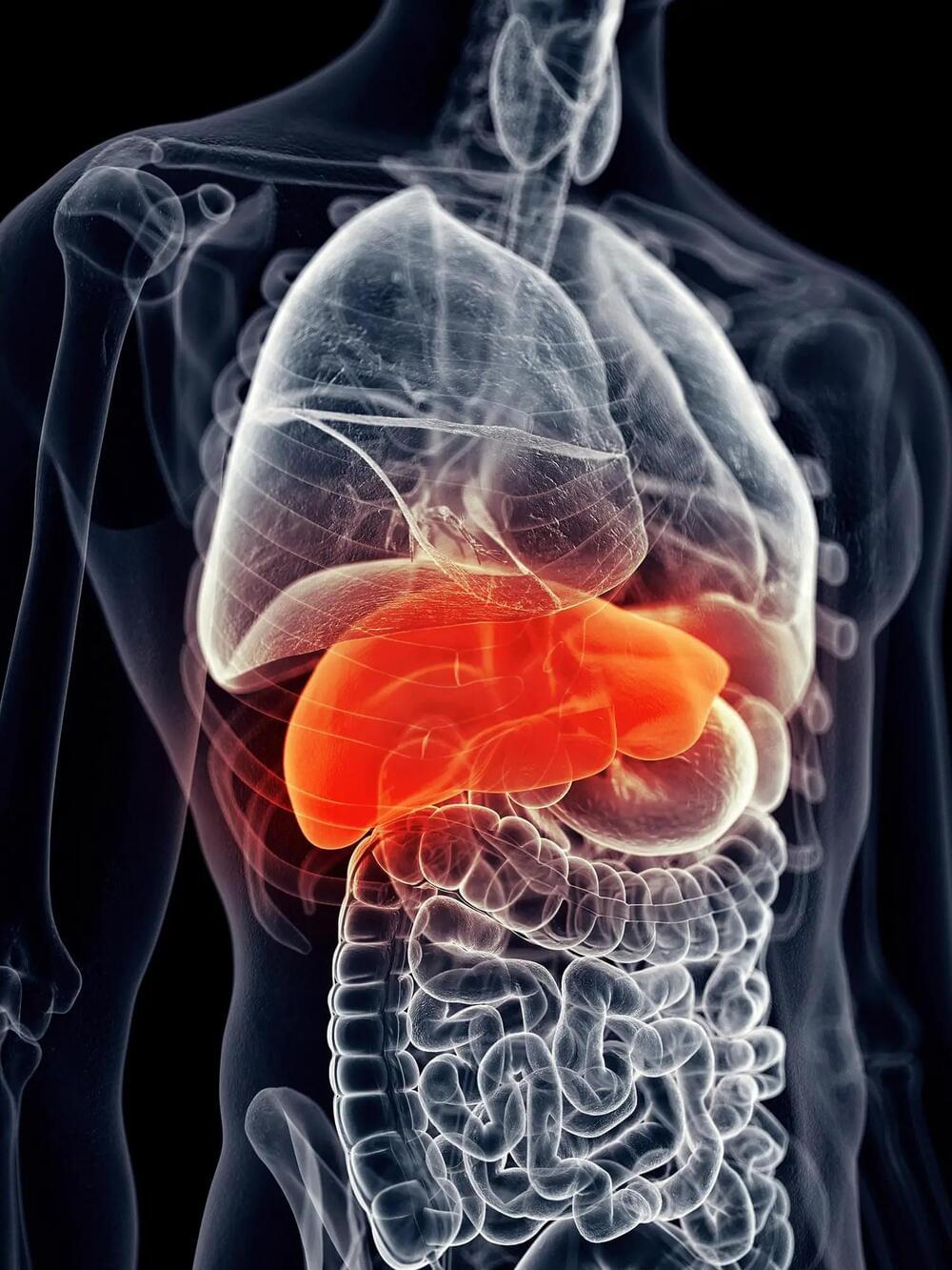
A new study from Sanford Burnham Prebys has discovered a drug that can spur liver regeneration in patients with Alagille syndrome.
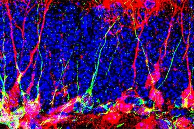
For the first time, research conducted by Associate Professor Duc Dong, Ph.D. has revealed that the detrimental effects of Alagille syndrome, a genetic disorder that has no cure, can be reversed using a single drug. The findings, published in the Proceedings of the National Academy of Sciences, have the potential to revolutionize the treatment approach for this rare condition, and could also shed light on more widespread diseases.
“Alagille syndrome is widely considered an incurable disease, but we believe we’re on the way to changing that,” says Dong, who is also the associate dean of admissions for Sanford Burnham Prebys’ graduate school. “We aim to advance this drug into clinical trials, and our results demonstrate its effectiveness for the first time.”