The integration of mechanical memory in the form of springs has for hundreds of years proven to be a key enabling technology for mechanical devices (such as clocks), achieving advanced functionality through complex autonomous movements. Currently, the integration of springs in silicon-based microtechnology has opened the world of planar mass-producible mechatronic devices from which we all benefit, via air-bag sensors for example.
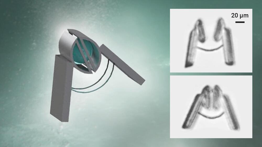
For a new generation of minimally and even non-invasive biomedical applications however, mobile devices that can safely interact mechanically with cells must be achieved at much smaller scales (10 microns) and with much softer forces (pico Newton scale, i.e., lifting weights less than one millionth of a mg) and in customized three-dimensional shapes.
Researchers at the Chemnitz University of Technology, the Shenzhen Institute of Advanced Technology of the Chinese Academy of Sciences and the Leibniz IFW Dresden, in a recent publication in Nature Nanotechnology, have demonstrated that controllable springs can be integrated at arbitrary chosen locations within soft three-dimensional structures using confocal photolithographic manufacturing (with nanoscale precision) of a novel magnetically active material in the form of a photoresist impregnated with customizable densities of magnetic nanoparticles.


 עברית (Hebrew)