Toggle light / dark theme

First pig-to-human cardiac transplant alters heart’s electrical signals

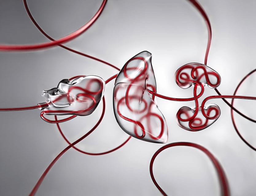
A recent study to be presented at the American Heart Association (AHA) Scientific Sessions 2022 revealed unexpected changes in the electrical conduction system of the first genetically-modified porcine-to-human heart xenotransplant.

Xenotransplantation is the procedure of transplantation/implantation into a human of organs from non-human animal sources. The first pig-to-human heart xenograft was transplanted in January 2022 at the University of Maryland. The recipient survived for 61 days after receiving the xenograft. Research efforts have been underway for this xenotransplantation for over three decades.

Harvesting genetically-modified porcine hearts, the genes of which have been altered for safe transplantation into humans, would become a reality if successful. However, xenotransplantation of organs into a human carries several inherent challenges. With these transplant procedures, there is always the risk of graft rejection, infection, and abnormal heart rhythms.

DNA ‘nanotransporters’ to treat cancer

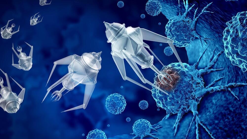
A team of Canadian researchers from Université de Montréal has designed and validated a new class of drug transporters made of DNA that are 20,000 times smaller than a human hair and that could improve how cancers and other diseases are treated.

Reported in a new study in Nature Communications, these molecular transporters can be chemically programmed to deliver optimal concentration of drugs, making them more efficient than current methods.

Communication Breakdown: Into the Molecular Mechanism of Biofilm Inhibition

Bacterial biofilm formation is a huge problem in industry and medicine. Therefore, the discovery of anti-biofilm agents may hold great promise. Biofilm formation is usually a consequence of bacterial cell–cell communication, a process called quorum sensing (QS). CeO2 nanocrystals (NCs) have been established as haloperoxidase (HPO) mimics and ecologically beneficial biofilm inhibitors. They were suggested to interfere with QS, a mechanism termed quorum quenching (QQ), but their molecular mechanism remained elusive. We show that CeO2 NCs are effective QQ agents, inactivating QS signals by bromination. Catalytic bromination of 3-oxo-C12-AHL a QS signaling compound used by Pseudomonas aeruginosa, was detected in the presence of CeO2 NCs, bromide ions, and hydrogen peroxide. Brominated acyl-homoserine lactones (AHLs) no longer act as QS signals but were not detected in the bacterial cultures.

All-Optical Modulation of Single Defects in Nanodiamonds: Revealing Rotational and Translational Motions in Cell Traction Force Fields

Measuring the mechanical interplay between cells and their surrounding microenvironment is vital in cell biology and disease diagnosis. Most current methods can only capture the translational motion of fiduciary markers in the deformed matrix, but their rotational motions are normally ignored. Here, by utilizing single nitrogen-vacancy (NV) centers in nanodiamonds (NDs) as fluorescent markers, we propose a linear polarization modulation (LPM) method to monitor in-plane rotational and translational motions of the substrate caused by cell traction forces. Specifically, precise orientation measurement and localization with background suppression were achieved via optical polarization selective excitation of single NV centers with precisions of ∼0.5°/7.5 s and 2 nm/min, respectively.

Dr. Jacob Hanna, MD, Ph.D. — Synthetic Embryo R&D In Regenerative Medicine & Developmental Biology

(https://hannalabweb.weizmann.ac.il/) is a Senior Scientist and Professor in the Department of Molecular Genetics at the Weizmann Institute of Science in Israel, where his lab, and the interdisciplinary group of scientists within it, are focused on understanding the complexity of early embryonic stem cell biology and early developmental dynamics, as well as advancing human disease modeling.

More specifically, Dr. Hanna’s lab investigates the detailed process of cellular reprogramming, in which induced pluripotent stem cells are generated from somatic cells, and they investigate how pluripotency is maintained throughout development in mouse and human. In their studies they employ a diverse arsenal of biological experimentation methods, high throughput screening, advanced microscopy and genomic analyses seeking to combine biological experimentation with computational biology, theory and modeling, to elucidate various biological questions.

Dr. Hanna completed both his MD and PhD at The Hebrew University of Jerusalem, where his work was focused in the domain of immunology with his thesis focus on novel molecular and functional properties of human NK Subsets. He then went on to do postdoctoral studies at the Whitehead Institute for Biomedical Research at MIT under the tutelage of Prof. Dr. Rudolf Jaenisch with a field of study of pluripotency and epigenetic reprogramming.

Dr. Hanna is also Founder and Chief Scientific Advisor, of Renewal Bio (https://www.renewal.bio/), a biotech company looking to leverage the power of these new stem cell technologies, potentially applying them to a wide variety of human ailments including infertility, genetic diseases, and longevity.

Sites in the brain where RNA is edited could help our understanding of neurodevelopment and disease

Mount Sinai researchers have cataloged thousands of sites in the brain where RNA is modified throughout the human lifespan in a process known as adenosine-to-inosine (A-to-I) editing, offering important new avenues for understanding the cellular and molecular mechanisms of brain development and how they factor into both health and disease.

In a study published in Cell Reports, the team described how the rate of RNA editing in the brain increases as individuals age, with implications for dissecting the pathology of altered A-to-I editing across a range of neurodevelopmental and aging disorders.

“Our work provides more nuanced and accurate insights into the contribution of RNA modifications by A-to-I editing during human brain development,” says senior author Michael Breen, Ph.D., Assistant Professor of Psychiatry, and Genetics and Genomic Sciences, at the Icahn School of Medicine at Mount Sinai, and a member of the Seaver Center for Autism Research and Treatment.

Anthropologists find new ways female bones are permanently altered after giving birth

Reproduction permanently alters females’ bones in ways not previously known, a team of anthropologists has found. Its discovery, based on an analysis of primates, sheds new light on how giving birth can permanently change the body.

“Our findings provide additional evidence of the profound impact that reproduction has on the female organism, further demonstrating that the skeleton is not a static organ, but a dynamic one that changes with ,” explains Paola Cerrito, who led the research as a doctoral student in NYU’s Department of Anthropology and College of Dentistry.

Specifically, the researchers found that calcium, magnesium, and are lower in females who have experienced reproduction. These changes are linked to giving birth itself and to lactation.

New research rethinks the blood-tumor barrier and identifies novel path to brain cancer treatment

In a new study, scientists have uncovered the mechanics of the blood-tumor barrier, one of the most significant obstacles to improving treatment efficacy and preventing the return of cancerous cells. The research team, led by Dr. Xi Huang, a Senior Scientist in Developmental & Stem Cell Biology program at The Hospital for Sick Children (SickKids), lays the foundation for more effectively treating medulloblastoma, the most common malignant pediatric brain tumor.

“Despite decades of research on brain cancer, the mechanisms that govern the formation and function of the blood-tumor barrier have remained poorly understood,” says Huang, who is also a Principal Investigator at the Arthur and Sonia Labatt Brain Tumor Research Center and Canada Research Chair in Cancer Biophysics. “Our discoveries represent a breakthrough in the understanding of how the blood-tumor barrier forms and works.”

In a paper published today in Neuron, the research team identifies a way to reduce the impact of the blood-tumor barrier on medulloblastoma treatment.

Using sound to model the world

Imagine the booming chords from a pipe organ echoing through the cavernous sanctuary of a massive, stone cathedral.

The a cathedral-goer will hear is affected by many factors, including the location of the organ, where the listener is standing, whether any columns, pews, or other obstacles stand between them, what the walls are made of, the locations of windows or doorways, etc. Hearing a sound can help someone envision their environment.

Researchers at MIT and the MIT-IBM Watson AI Lab are exploring the use of spatial acoustic information to help machines better envision their environments, too. They developed a that can capture how any sound in a room will propagate through the space, enabling the model to simulate what a listener would hear at different locations.