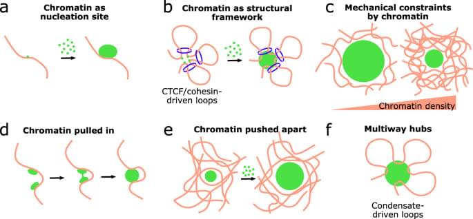
In this Mini Review, the authors discuss the relationship between condensate formation, genome organization, and transcriptional activity, focusing on the experimental evidence behind the role of transcriptional condensates in gene regulation.
Category: biotech/medical – Page 1,263

Endovascular thrombectomy found to be beneficial for large ischemic stroke
For patients with acute ischemic stroke and large cores, endovascular thrombectomy (EVT) improves clinical outcomes compared with medical management (MM), according to a study published online Feb. 7 in the Journal of the American Medical Association to coincide with the annual American Stroke Association International Stroke Conference, which was held from Feb. 7 to 9 in Phoenix.
Amrou Sarraj, M.D., from Case Western Reserve University in Cleveland, and colleagues describe the relationship between imaging estimates of irreversibly injured brain and at-risk regions and clinical outcomes and EVT treatment effect in an exploratory analysis of the SELECT2 trial.
Adults with acute ischemic stroke due to occlusion of the internal carotid or middle cerebral artery (M1 segment) and large ischemic core were randomly allocated to EVT versus MM across 31 global centers; the analysis included 336 patients.
The Limits of Immortality & Digital Death
It is often thought that if we cure aging or find out how to upload a human mind that humans will be immortal. Today we will examine that notion and see how well it holds up against astronomical time lines.
Visit our Website: http://www.isaacarthur.net.
Join Nebula: https://go.nebula.tv/isaacarthur.
Support us on Patreon: / isaacarthur.
Support us on Subscribestar: https://www.subscribestar.com/isaac-a…
Facebook Group: / 1583992725237264
Reddit: / isaacarthur.
Twitter: / isaac_a_arthur on Twitter and RT our future content.
SFIA Discord Server: / discord or Download the audio of this episode from Soundcloud: / digital-death.
Cover Art by Jakub Grygier: https://www.artstation.com/artist/jak…
Graphics Team:
Edward Nardella.
Jarred Eagley.
Justin Dixon.
Katie Byrne.
Kris Holland of Mafic Stufios: www.maficstudios.com.
Misho Yordanov.
Pierre Demet.
Sergio Botero: https://www.artstation.com/sboterod?f…
Stefan Blandin.
Script Editing:
Andy Popescu.
Connor Hogan.
Edward Nardella.
Eustratius Graham.
Gregory Leal.
Jefferson Eagley.
Luca de Rosa.
Mark Warburton.
Michael Gusevsky.
Mitch Armstrong.
MolbOrg.
Naomi Kern.
Philip Baldock.
Sigmund Kopperud.
Steve Cardon.
Tiffany Penner.
Music:
Markus Junnikkala, \
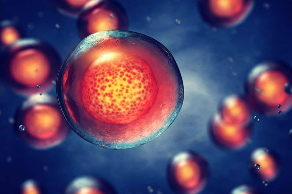
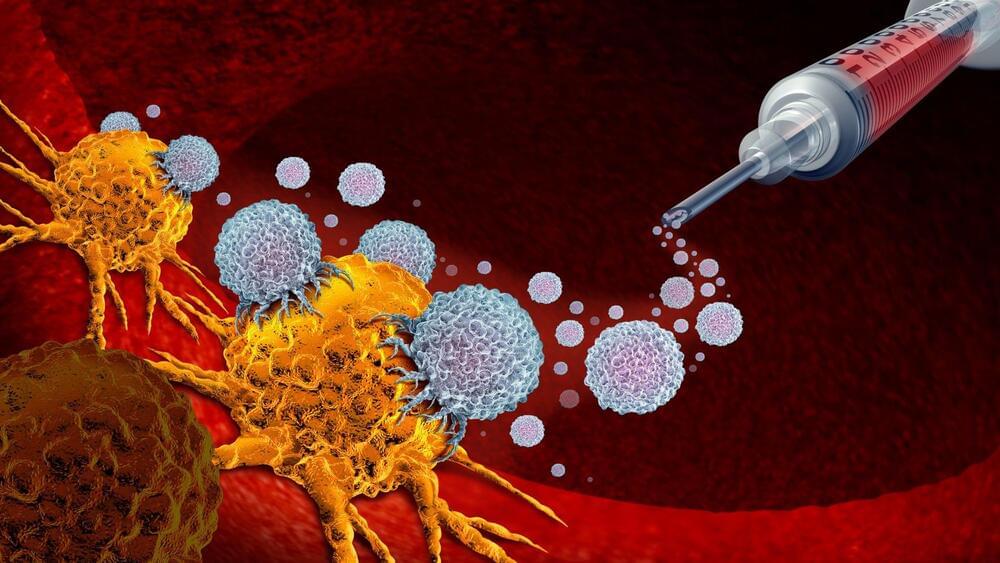
Stem cells grown in labs for experimental therapies pose a cancer risk
Around one-fifth of the stem cells grown in laboratories for as-yet-unapproved medical treatments have cancer-causing mutations.
By Clare Wilson
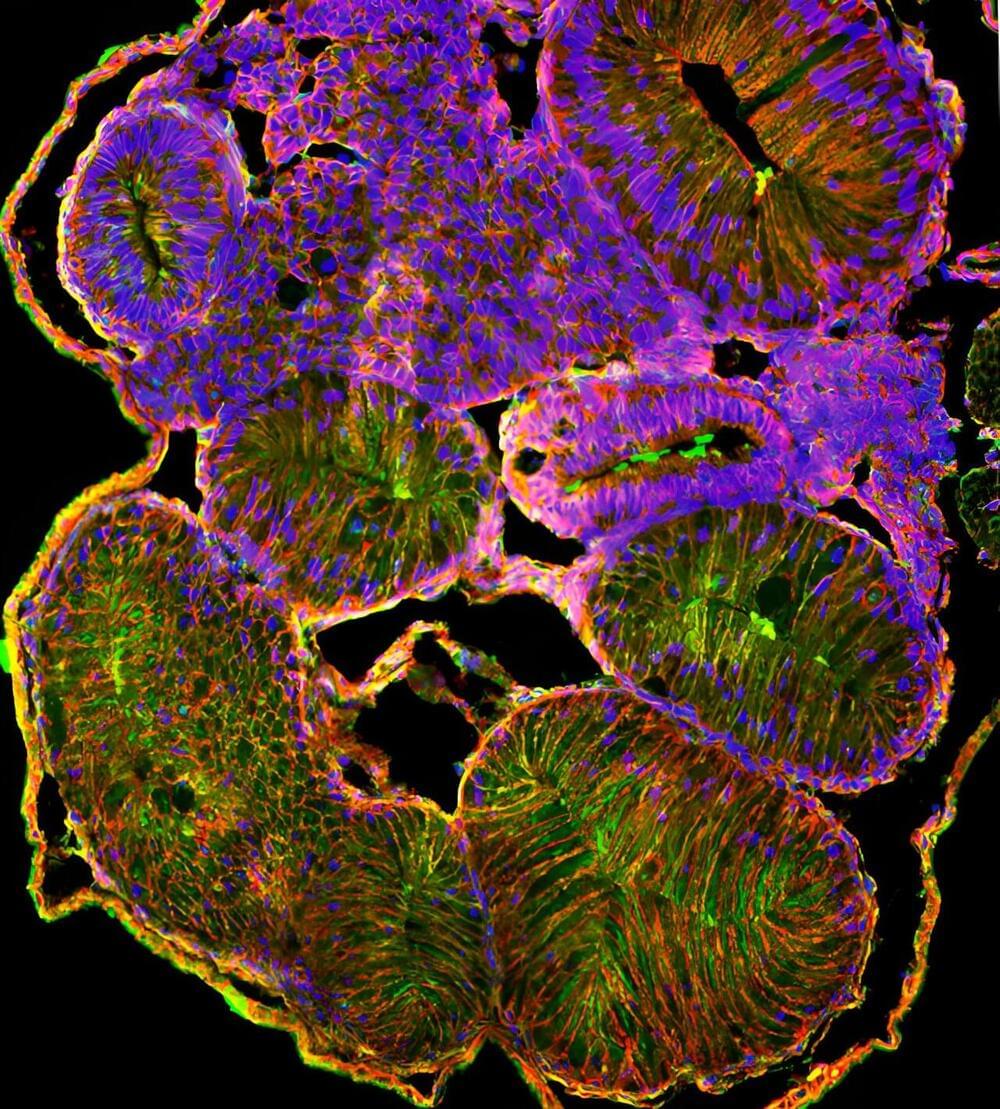
Frog embryo study helps scientists unravel the human birth anomaly of intestinal malrotation
How does our intestine, which can be at least 15 feet long, fit properly inside our bodies? As our digestive system grows, the gut tube goes through a series of dramatic looping and rotation to package the lengthening intestine. Failure of the gut to rotate properly during development results in a prevalent, but poorly understood, birth anomaly called intestinal malrotation.
Now, in a study published in the journal Development, scientists from North Carolina State University have uncovered a potential cause of this life-threatening condition.
Intestinal malrotation affects 1 in 500 births but the underlying causes are not well understood. To find out why gut revolution could go amiss, scientists need to first understand intestinal rotation during normal development, a complex process that still baffles biologists.
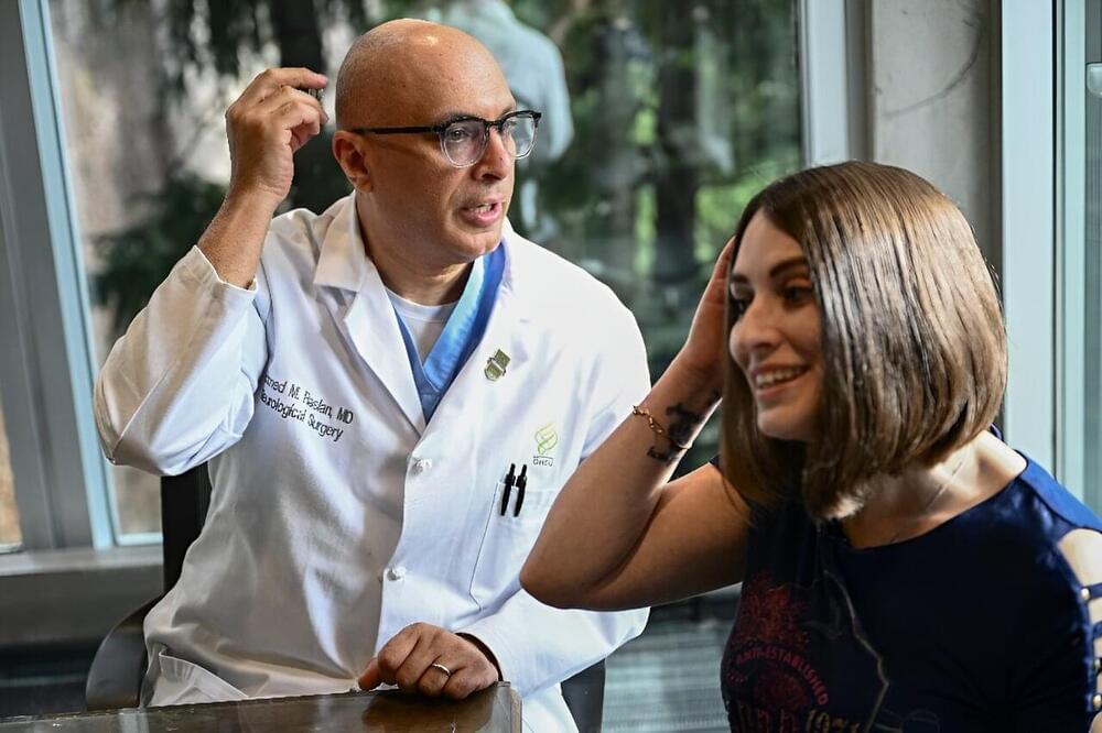
US Patient ‘Happy Again’ after Brain Implant Treats Epilepsy and OCD
American Amber Pearson used to wash her hands until they bled, terrified by the idea of contamination from everyday items, a debilitating result of her obsessive compulsive disorder (OCD).
But the repetitive rituals of her condition are largely consigned to memory, thanks to a revolutionary brain implant that is being used to treat both her epilepsy and her OCD.
“I’m actually present in my daily life and that’s incredible,” the 34-year-old told AFP.
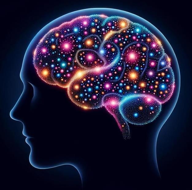
Important Implications: New Study Aims To Unlock Secrets of the Human Brain
Dr. Richard Naud’s research at the University of Ottawa holds important implications for understanding learning and memory theories, and it may pave the way for advancements in artificial intelligence in the future.
The mysteries of the human brain’s internal mechanisms are slowly being uncovered, and a significant new study led by Dr. Richard Naud from the Faculty of Medicine at the University of Ottawa is bringing us nearer to solving these profound questions.
The study’s results have important implications for theories of learning and working memory and could potentially help lead to future developments in artificial intelligence (AI) since AI developers and programmers watch the work of Dr. Naud and other leading neuroscientists.

Global project to drive lifesaving agreement on appropriate antimicrobial drug use
University of Melbourne researchers are leading a new push to address the growing harm of antimicrobial resistance (AMR) as more humans and animals become seriously ill or die from infections that medicine once treated easily.
Over-use and misuse of microbe-killing drugs – including antibiotics, antivirals and antifungals – is the main driver accelerating the evolution of resistance to these drugs in bacteria, viruses, fungi and parasites around the world.
The World Health Organisation calls AMR a top global public health threat that was directly responsible for 1.27 million deaths and contributed to 4.95 million deaths in 2019.