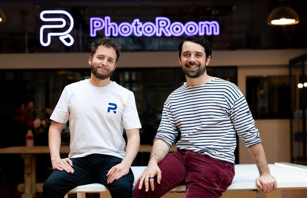
Toggle light / dark theme

The microbots are applied nasally to treat brain diseases.


Scientists have successfully guided a microbot through the nasal pathways to the brain of a mouse. If the same approach can be replicated in humans, it could be a game-changer against neurodegenerative disease, enabling doctors to deliver therapies directly to the brain.

A research team led by DGIST (the Daegu Gyeongbuk Institute of Science and Technology in South Korea) has created a microrobot propelled by magnets that can navigate the human body. The trial, published in the journal Advanced Materials, describes how they manufactured the microrobot, dubbed a Cellbot, by magnetizing stem cells extracted from the human nasal cavity. The scientists then tested the ability of the Cellbot to move through the body’s confined vessels and passages to reach its target, which it completed with ease.

DGIST said in a statement that “This approach has the potential to effectively treat central nervous system disorders in a minimally invasive manner.”

A robot has performed laparoscopic surgery on the soft tissue of a pig without the guiding hand of a human—a significant step in robotics toward fully automated surgery on humans. Designed by a team of Johns Hopkins University researchers, the Smart Tissue Autonomous Robot (STAR) is described today in Science Robotics.

“Our findings show that we can automate one of the most intricate and delicate tasks in surgery: the reconnection of two ends of an intestine. The STAR performed the procedure in four animals and it produced significantly better results than humans performing the same procedure,” said senior author Axel Krieger, an assistant professor of mechanical engineering at Johns Hopkins’ Whiting School of Engineering.

The robot excelled at intestinal anastomosis, a procedure that requires a high level of repetitive motion and precision. Connecting two ends of an intestine is arguably the most challenging step in gastrointestinal surgery, requiring a surgeon to suture with high accuracy and consistency. Even the slightest hand tremor or misplaced stitch can result in a leak that could have catastrophic complications for the patient.

“The change in life expectancy is significant, when you consider that an equivalent jump in human life expectancy would have us living on average until almost 120,” lead researcher Haim Cohen of Bar-Ilan University told the Times of Israel.

A longer life: The average human life expectancy has doubled in just the past 200 years, thanks in no small part to scientific breakthroughs in medicine, nutrition, and disease.

A team of researchers from the University of Arizona has proposed a “Lunar Ark” for preserving samples of 6.7 million Earth species in the event of a global crisis.

Be the first to know about the latest updates on COVID-19 pandemic, lockdowns, community quarantine, new normal, and Serbisyong Bayanihan.

We Serve the People. We Give Glory To God!
#SerbisyongBayanihan #UNTVNewsandRescue #LagingHandaPH

For news update, visit: https://www.untvweb.com/news/

Today, the ECoG grids most commonly used in surgeries typically have between 16 and 64 sensors, although research grade grids with up to 256 sensors can be custom made. The device created at UCSD is therefore a major advance in the field. It could improve neurosurgeons’ ability to remove as much of a brain tumour as possible while minimising damage to healthy tissue. In the case of epilepsy, the higher resolution could enable a surgeon to precisely identify the brain regions where seizures are originating, so that these can be removed without touching nearby regions not involved in seizure initiation. In this way, these high-resolution grids may enhance preservation of normal, functioning brain tissue.

ECoG grids with sensors in the thousands could also help in uncovering a deeper understanding of how the brain functions. Basic science advances, in turn, could lead to improved treatments grounded in enhanced understanding of brain function.

The team at UCSD – who collaborated with Massachusetts General Hospital and Oregon Health & Science University – achieved their breakthrough by packing individual sensors significantly closer to each other, while avoiding problematic interference between nearby sensors. The ECoG grids already in clinical use typically have sensors that are spaced one centimetre apart. But the new 1,024-sensor device has sensors just one millimetre apart, with a total grid area measuring three by three centimetres and is scalable to 2,048 sensors.

Protective coatings are common for many things in daily life that see a lot of use. We coat wood floors with finish; apply Teflon to the paint on cars; even use diamond coatings on medical devices. Protective coatings are also essential in many demanding research and industrial applications.

Now, researchers at Los Alamos National Laboratory have developed and tested an atomically thin coating for next-generation, electron-beam accelerator equipment—perhaps the most challenging technical application of the technology, the success of which bears out the potential for “Atomic Armor” in a range of applications.

“Accelerators are important tools for addressing some of the faced by humanity,” said Hisato Yamaguchi, member of the Sigma-2 group at the Laboratory. “Those challenges include the quest for , continued scaling of computational power, detection and mitigation of pathogens, and study of the structure and dynamics of the building blocks of life. And those challenges all require the ability to access, observe and control matter on the frontier timescale of electronic motion and the spatial scale of atomic bonds.”

Scanning for Memories

At the time there was almost no evidence of this from neuron studies. But in 2006, Ma, Pouget and their colleagues at the University of Rochester presented strong evidence that populations of simulated neurons could perform optimal Bayesian inference calculations. Further work by Ma and other researchers over the past dozen years offered additional confirmations from electrophysiology and neuroimaging that the theory applies to vision by using machine learning programs called Bayesian decoders to analyze actual neural activity.

Neuroscientists have used decoders to predict what people are looking at from fMRI (functional magnetic resonance imaging) scans of their brains. The programs can be trained to find the links between a presented image and the pattern of blood flow and neural activity in the brain that results when people see it. Instead of making a single guess — that the subject is looking at an 85-degree angle, for instance — Bayesian decoders produce a probability distribution. The mean of the distribution represents the likeliest prediction of what the subject is looking at. The standard deviation, which describes the width of the distribution, is thought to reflect the subject’s uncertainty about the sight (is it 85 degrees or could it be 84 or 86?).

A team of engineers and neurosurgeons developed a state-of-the-art brain sensor that could greatly enhance the treatment of cancer and epilepsy, according to a press statement from the University of California San Diego.

The new apparatus can record electrical signals from the brain’s surface in a never-before-seen resolution for such a device.

The breakthrough opens up new possibilities for brain-computer interfaces, such as the ones being developed by Elon Musk’s Neuralink. Not only will this help to improve diagnoses of deadly diseases, it also has the potential to transform our understanding of the human brain.

And it took less than a full workday. Stanford Medicine scientists and their collaborators have engineered a new genome sequencing technique that can diagnose rare genetic diseases in an average of eight hours. This is a record-breaking time frame that is leap and bounds ahead of other current advanced technologies.


Gene sequencing is crucial to advancing science! Check out why cutting time and cost is key.

Researchers from the University of Birmingham and the University of Huddersfield, UK, have developed a new 3D bioprinting technique that can be used to treat chronic wounds.

Named Suspended Layer Additive Manufacturing (SLAM), the approach enables the printing of a novel biomaterial that accurately simulates the structure of mammalian skin.

In fact, according to the researchers, the biomaterial is the first of its kind to simulate all three of the major layers found in skin – the hypodermis, the dermis, and the epidermis – making it a unique tri-layered skin equivalent. Early experiments suggest that the 3D bioprinted skin can be placed at the site of a wound to induce healing, reducing scar tissue in the process.