Some of the AI-designed gene editors could be more versatile than those found in nature.

Researchers at Umeå University, Sweden, have discovered that how a special protein complex called the Mediator moves along genes in DNA may have an impact on how cells divide. The discovery may be important for future research into the treatment of certain diseases. The study is published in Nucleic Acids Research.


As the US grapples with an ongoing bird flu outbreak in dairy cattle, the country’s health agencies are ramping up surveillance efforts and working to develop a vaccine if needed.
By Grace Wade

A research team at the University of Pittsburgh led by Alexander Star, a chemistry professor in the Kenneth P. Dietrich School of Arts and Sciences, has developed a fentanyl sensor that is six orders of magnitude more sensitive than any electrochemical sensor for the drug reported in the past five years. The portable sensor can also tell the difference between fentanyl and other opioids.
Hvidovre Hospital has the world’s first prototype of a sensor capable of detecting errors in MRI scans using laser light and gas. The new sensor, developed by a young researcher at the University of Copenhagen and Hvidovre Hospital, can thereby do what is impossible for current electrical sensors—and hopefully pave the way for MRI scans that are better, cheaper and faster.

The following is a summary of “Comparative Effectiveness of Partial Gland Cryoablation Versus Robotic Radical Prostatectomy for Cancer Control,” published in the April 2024 issue of Urology by Zhu et al.
In this study, researchers address the notable gap in high-level evidence comparing oncologic endpoints for partial gland ablation, where most existing series rely on prostate-specific antigen (PSA) rather than biopsy endpoints. The objective was to conduct a comprehensive comparison of oncologic outcomes between partial gland cryoablation (PGC) and radical prostatectomy (RP) for the management of prostate cancer.
Through a retrospective, single-center analysis, investigators examined a cohort of subjects treated with either PGC (n = 98) or RP (n = 536) as primary treatment for intermediate-risk (Gleason grade group [GG] 2–3) prostate cancer between January 2017 and December 2022. Key oncologic endpoints included surveillance biopsies per protocol after PGC and serial PSA testing after RP. The primary outcome of interest was treatment failure, which is defined as the necessity for salvage treatment or metastatic disease development. The study group conducted treatment failure and survival analyses using Cox proportional-hazard regression and Kaplan-Meier survival curves. After carefully applying inclusion/exclusion criteria, they compared the PGC (n = 75) and RP (n = 298) groups.


In Limitless, the film’s protagonist is a struggling writer desperate for success. As luck would have it, he is introduced to an experimental drug that unlocks his full potential.
The film was a box office success, and it is easy to see why.
Nootropics, drugs, and various supplements for memory, focus, and mental agility have been used for millennia. The desire to elevate ourselves is universal, much like the quest for longevity–and, unsurprisingly, the two are intertwined.
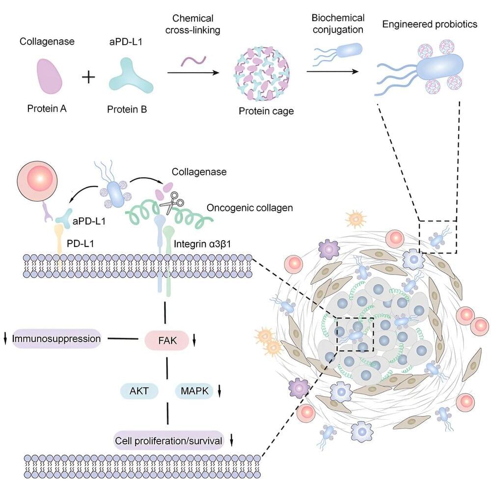
Many pancreatic tumors are like malignant fortresses, surrounded by a dense matrix of collagen and other tissue that shields them from immune cells and immunotherapies that have been effective in treating other cancers. Employing bacteria to infiltrate that cancerous fortification and deliver these drugs could aid treatment for pancreatic cancer, according to newly published findings from a team of University of Wisconsin–Madison researchers.