Toggle light / dark theme

Dr. Behnaam Aazhang, Ph.D. — Director, Rice Neuroengineering Initiative (NEI), Rice University

Restoring And Extending The Capabilities Of The Human Brain — Dr. Behnaam Aazhang, Ph.D. — Director, Rice Neuroengineering Initiative, Rice University


Dr. Behnaam Aazhang, Ph.D. (https://aaz.rice.edu/) is the J.S. Abercrombie Professor, Electrical and Computer Engineering, and Director, Rice Neuroengineering Initiative (NEI — https://neuroengineering.rice.edu/), Rice University, where he has broad research interests including signal and data processing, information theory, dynamical systems, and their applications to neuro-engineering, with focus areas in (i) understanding neuronal circuits connectivity and the impact of learning on connectivity, (ii) developing minimally invasive and non-invasive real-time closed-loop stimulation of neuronal systems to mitigate disorders such as epilepsy, Parkinson, depression, obesity, and mild traumatic brain injury, (iii) developing a patient-specific multisite wireless monitoring and pacing system with temporal and spatial precision to restore the healthy function of a diseased heart, and (iv) developing algorithms to detect, predict, and prevent security breaches in cloud computing and storage systems.

Dr. Aazhang received his B.S. (with highest honors), M.S., and Ph.D. degrees in Electrical and Computer Engineering from University of Illinois at Urbana-Champaign in 1981, 1983, and 1986, respectively. From 1981 to 1985, he was a Research Assistant in the Coordinated Science Laboratory, University of Illinois. In August 1985, he joined the faculty of Rice University. From 2006 till 2014, he held an Academy of Finland Distinguished Visiting Professorship appointment (FiDiPro) at the University of Oulu, Oulu, Finland.

Dr. Aazhang is a Fellow of IEEE and AAAS, and a distinguished lecturer of IEEE Communication Society.

Dr. Aazhang received an Honorary Doctorate degree from the University of Oulu, Finland (the highest honor that the university can bestow) in 2017 and IEEE ComSoc CTTC Outstanding Service Award “For innovative leadership that elevated the success of the Communication Theory Workshop” in 2016. He is a recipient of 2004 IEEE Communication Society’s Stephen O. Rice best paper award for a paper with A. Sendonaris and E. Erkip. In addition, Sendonaris, Erkip, and Aazhang received IEEE Communication Society’s 2013 Advances in Communication Award for the same paper. He has been listed in the Thomson-ISI Highly Cited Researchers and has been keynote and plenary speaker of several conferences.
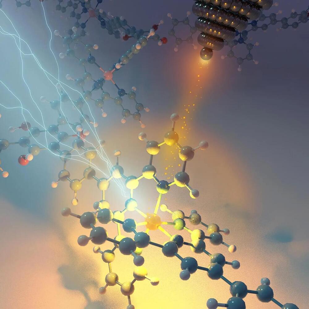
For the Very First Time: Scientists Analyze a Single Atom With X-Rays

New X-ray capability could find wide application in environmental and medical research, as well as the development of batteries and microelectronic devices.

Scientists at Argonne National Laboratory and various universities have successfully used X-ray beams to analyze a single atom.

An atom is the smallest component of an element. It is made up of protons and neutrons within the nucleus, and electrons circling the nucleus.

Colorectal cancer in the liver: New treatment gives more people hope for a cure

Health care professionals are overcoming these obstacles with a new treatment called hepatic artery infusion pump chemotherapy that shrinks liver tumors, giving more people a chance for surgery. This treatment also can shrink tumors in the bile ducts inside the liver, called intrahepatic cholangiocarcinoma.

“Our goal is to expand the number of patients who could be offered curative treatment,” says Dr. Thiels. “We are also aiming to reduce the risk of cancer recurring in people with high-risk liver tumors.”

The hepatic artery carries oxygen-rich blood to the liver. A hepatic artery pump delivers chemotherapy directly into the liver’s blood supply. Because chemotherapy from the pump only reaches the liver, the approach can use higher doses to shrink liver tumors more effectively. “We can often achieve a higher response rate than conventional chemotherapy,” says Dr. Thiels.

New biomarker confirmed for early diagnosis of multiple sclerosis

A study conducted by researchers from the Department of Neurology at MedUni Vienna and University Hospital Vienna has demonstrated for the first time that the diagnosis of multiple sclerosis (MS) can be significantly improved by additionally measuring the thickness of retinal layers in the eye.

Use of the procedure, which is already available at the Departments of MedUni Vienna and University Hospital Vienna, helps to detect the condition at an earlier stage and predict its progression more accurately. This can lead to a decisive increase in the chance of improved patient outcomes. The findings have been published in the journal Neurology.

As part of their investigation, the research team headed by Gabriel Bsteh and Thomas Berger of the Department of Neurology at MedUni Vienna and University Hospital Vienna collaborated with colleagues from MedUni Vienna and University Hospital Vienna’s Department of Ophthalmology and Optometrics to examine 267 MS patients over a period of five years.

Living digital camera: scientists capture light patterns in DNA for image storage

In a recent article published in Nature communications*, researchers described a technique of capturing two-dimensional (2D) light patterns into deoxyribonucleic acid (DNA) and using high-throughput next-generation sequencing to retrieve recorded images.

Study: A biological camera that captures and stores images directly into DNA. Image Credit: BillionPhotos/Shutterstock.com.

Smile, You’re on BacCam! DNA Stores Images from a Living Digital Camera

Scientists at the National University of Singapore (NUS) have used bacteria for recording, storing, and retrieving images in DNA. This biological analog to a digital camera, which the authors have named “BacCam,” is a crucial step for DNA data storage techniques and the merging of biological and electronic systems.

The article, “A biological camera that captures and stores images directly into DNA,” was published in Nature Communications.

Prior to this publication, there were two landmark papers that addressed either the use of cells to capture light or the storage of images into DNA, but not the two together. In May 2017, researchers from the lab of Christopher Voigt, PhD, at the Massachusetts Institute of Technology (MIT) developed a system to produce ‘color photographs’ on bacterial culture plates by controlling pigment production and to redirect metabolic flux by expressing CRISPRi guide RNAs. Two months later, researchers in the lab of George Church, PhD, at Harvard Medical School demonstrated a method for encoding images via de novo DNA synthesis before insertion into the bacterial genome.

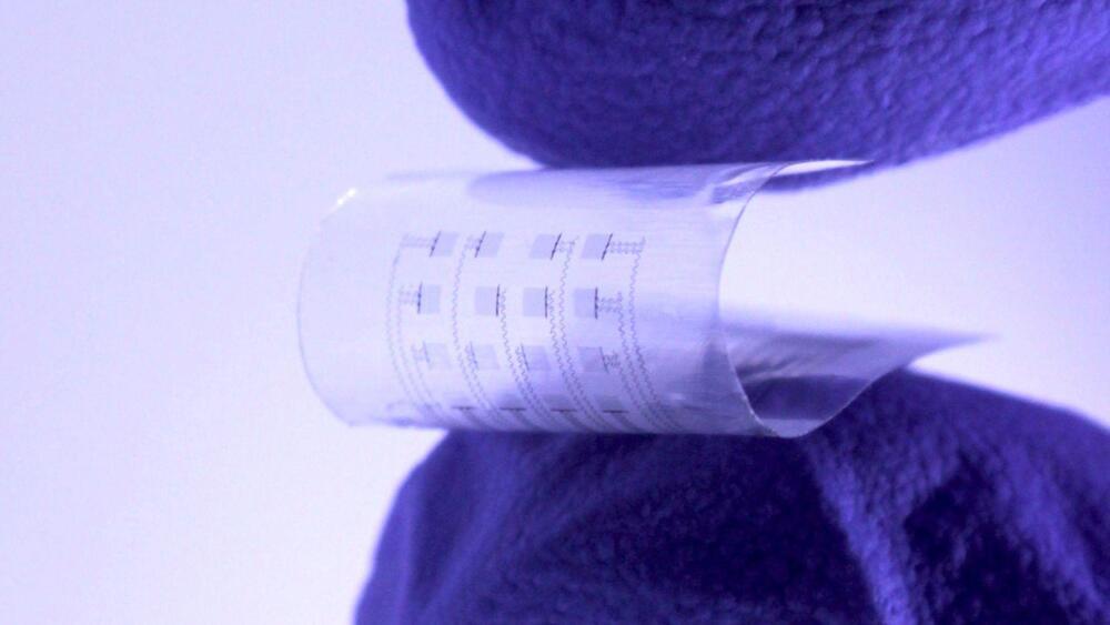
Dissolving electronic device to monitor and treat heart dysfunction gets FDA approval

New gadget can map electrical activity and deliver electrical stimuli to stop atrial fibrillation, before dissolving into the body.

Nearly 800K people fall victim to a heart attack in the US each year. A recent study undertaken by a team of researchers at Northwestern and George Washington (GW) universities has developed a new device to monitor and treat heart disease and dysfunctions in the aftermath of heart-related incidents.

The device seems to hold promise for providing critical support during the days, weeks, or months following heart problems. According to the researchers convey that the new technology harmlessly dissolves inside the body and skips the need for extraction.


Northwestern university / george washington university.

A recent study undertaken by a team of researchers at Northwestern and George Washington (GW) universities has developed a new device to monitor and treat heart disease and dysfunctions in the aftermath of heart-related incidents.

/* */