Synthego offers Full Stack Genome Engineering Solutions. Our Engineered Cells and CRISPR kits enables all researchers to access CRISPR and accelerate their scientific discoveries, uncover cures for diseases, and develop novel synthetic biology applications.
DARPA, the Department of Defense’s research arm, is paying scientists to invent ways to instantly read soldiers’ minds using tools like genetic engineering of the human brain, nanotechnology and infrared beams. The end goal? Thought-controlled weapons, like swarms of drones that someone sends to the skies with a single thought or the ability to beam images from one brain to another.
This week, DARPA (Defense Advanced Research Projects Agency) announced that six teams will receive funding under the Next-Generation Nonsurgical Neurotechnology (N3) program. Participants are tasked with developing technology that will provide a two-way channel for rapid and seamless communication between the human brain and machines without requiring surgery.
“Imagine someone who’s operating a drone or someone who might be analyzing a lot of data,” said Jacob Robinson, an assistant professor of bioengineering at Rice University, who is leading one of the teams. [DARPA’s 10 Coolest Projects: From Humanoid Robots to Flying Cars].
It’s a remarkable story of recovery, but it’s unclear how useful this sort of therapy could become.
The background: Isabelle Holdaway had been given less than a 1% chance of survival after a lung transplant, carried out to combat the symptoms of cystic fibrosis, left her with an antibiotic-resistant infection. She had been sent home and was in a terrible physical condition: underweight, with liver failure, and with lesions on her skin from the infection.
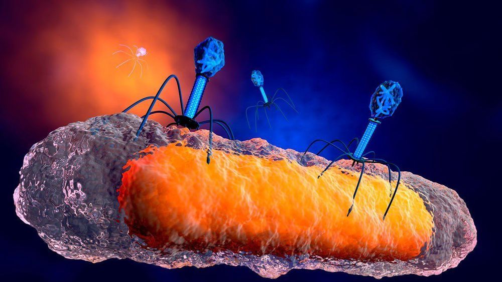
A breakthrough: Her consultant at Great Ormond Street Hospital in London worked with a team at the University of Pittsburgh to develop an untested phage therapy. This treatment used a cocktail of three phages, which are viruses that solely attack and kill bacteria. Two of the three phages, selected from a library of more more than 10,000 kept at the University of Pittsburgh, had been genetically engineered to be better at attacking the bacteria. The therapy was injected into her bloodstream twice daily and applied to the lesions on her skin, according to Nature Medicine.
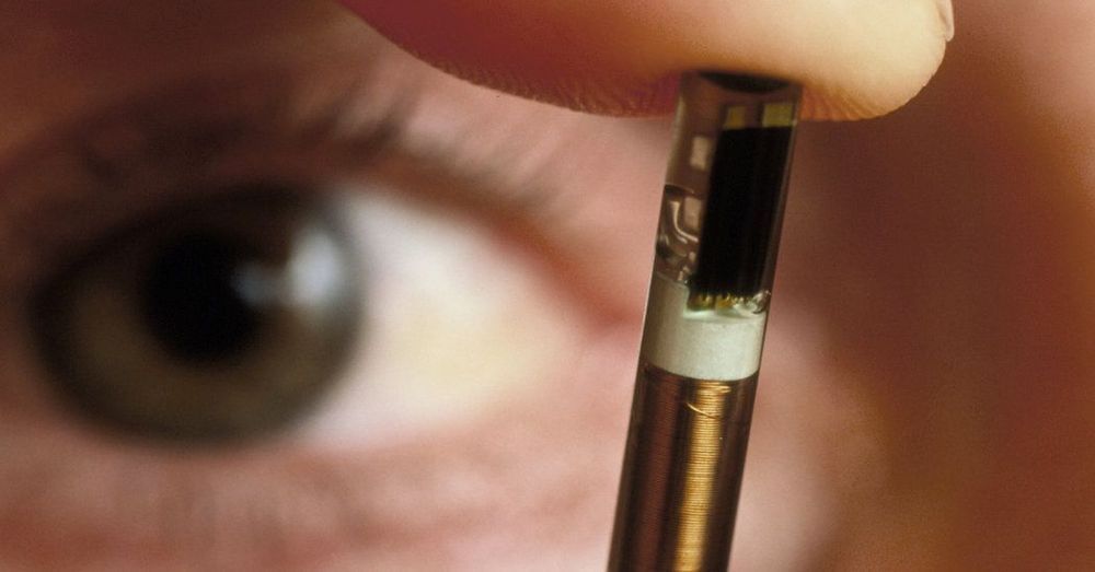
Nearly 50 years ago, The New York Times—widely considered America’s paper of record—changed the media industry by creating the first modern Op-Ed page. Since then, their Opinion section has arguably become the most important voice for many public ideas that enter and change the world. Everyone from Heads of State to the globe’s most powerful business people to Nobel Prize winners to everyday citizens have written there when they had something essential to say about the times we live in. I’m super excited to share my first Op-Ed for The New York Times on #biohacking and the growing concern of legalizing implants. It’s a happy professional day for me, and an important step forward for the growing #transhumanism movement as we begin to enter mainstream culture.
Implant technology can change the world — unless politicians give in to the hysteria against it.
Standard snapshots from space don’t quite show Earth in all its glory. There’s so much more to see.
To reveal details impossible to observe with the naked eye, Rice University engineers are building a portable spectrometer that can be mounted on a small satellite, flown on an airplane or a drone or someday even held in the hand.
Bioengineer Tomasz Tkaczyk and his colleagues at Rice’s Brown School of Engineering and Wiess School of Natural Sciences have published the first results from a NASA-funded project to develop a small, sophisticated spectrometer with unusual versatility. Their paper appears in Optics Express.
To answer the iconic question “Are We Alone?”, scientists around the world are also attempting to understand the origin of life. There are many pieces to the puzzle of how life began and many ways to put them together into a big picture. Some of the pieces are firmly established by the laws of chemistry and physics. Others are conjectures about what Earth was like four billion years ago, based on extrapolations of what we know from observing Earth today. However, there are still major gaps in our knowledge and these are necessarily filled in by best guesses.
We invited talented scientists to discuss their different opinions about the origin of life and the site of life’s origin. Most of them will agree that liquid water was necessary, but if we had a time machine and went back in time, would we find life first in a hydrothermal submarine setting in sea water or a fresh water site associated with emerging land masses?
Biologist David Deamer, a Research Professor of Biomolecular Engineering at the University of California, Santa Cruz, and multi-disciplinary scientist Bruce Damer, Associate Researcher in the Department of Biomolecular Engineering at UC Santa Cruz, will describe their most recent work, which infers that hydrothermal pools are the most plausible site for the origin of life. Both biologists have been collaborating since 2016 on a full conception of the Terrestrial Origin of Life Hypothesis.
Lynn Rothschild, Senior Scientist at NASA’s Ames Research Center and Adjunct Professor of Molecular Biology, Cell Biology, and Biochemistry at Brown University, who is an astrobiologist/ synthetic biologist specializing in molecular approaches to evolution, particularly in microbes and the application of synthetic biology to NASA’s missions, will provide an evolutionary biologist’s perspective on the subject.
Peter voss is a serial entrepreneur, engineer, inventor and a pioneer in artificial intelligence.
Peter started out in electronics engineering but quickly moved into software. After developing a comprehensive ERP software package, Peter took his first software company from a zero to 400-person IPO in seven years.
Fueled by the fragile nature of software, Peter embarked on a 20-year journey to study intelligence (how it develops in humans, how to measure it, and current AI efforts), and to replicate it in software. His research culminated in the creation of a natural language intelligence engine that can think, learn, and reason — and adapt to and grow with the user. He even coined the term ‘AGI’(Artificial General Intelligence) with fellow luminaries in the space.
Peter founded SmartAction.ai in 2009, which developed the first AGI-based call center automation technology. Now, in his latest venture, Aigo.ai, he is taking that technology a step further with the commercialization of the second generation of his ‘Conversational AI’ technology with a bold mission of providing hyper-intelligent hyper-personal assistants for everyone.
In addition to being an entrepreneur, engineer, inventor, and AI pioneer, Peter often writes and presents on various philosophical topics including rational ethics, free will, and artificial minds; and is deeply involved with futurism and radical life-extension.
Church of Perpetual Life, a science-based church is open to people of all faiths & belief systems. We are non-denominational & non-judgmental and a central gathering place of Transhumans. What unites us is our common faith, belief, and desire in Unlimited Life Spans.
In the past 24 hours, a story of potentially world-changing import has surfaced. First reported by the MIT Technology Review and then not long after by the Associated Press, who seem to have been sitting on the story for a while, the news that a Chinese scientist named He Jiankui led an unprecedented experiment to edit human embryos and see them carried to term rocked the genetics community. Here’s what you need to know about this evolving story.
The science
Besides He, the most important players in this story may be twin baby girls named Nana and Lulu. As far as we know the twins were edited as embryos using CRISPR-cas9, a gene editing tool. The stated purpose of the edit was to disable CCR5, a gene involved in allowing HIV to invade cells, which is how a virus infects a host.