126K likes, — ivyfusion on September 8, 2025



New brain research reveals why we’re willing to go out of our way to punish people who break the rules, even when it costs us time, money, or friends. This behavior, which researchers call “altruistic punishment,” has been essential for human cooperation since ancient times. It’s the invisible glue that keeps societies fair: we enforce the rules not just for ourselves, but for everyone.
Many people voluntarily incur costs to punish violations of social norms. Evolutionary models and empirical evidence indicate that such altruistic punishment has been a decisive force in the evolution of human cooperation. We used H2 15 O positron emission tomography to examine the neural basis for altruistic punishment of defectors in an economic exchange. Subjects could punish defection either symbolically or effectively. Symbolic punishment did not reduce the defector’s economic payoff, whereas effective punishment did reduce the payoff. We scanned the subjects’ brains while they learned about the defector’s abuse of trust and determined the punishment. Effective punishment, as compared with symbolic punishment, activated the dorsal striatum, which has been implicated in the processing of rewards that accrue as a result of goal-directed actions.

Cedars-Sinai researchers created “young” immune cells from human stem cells that reversed cognitive decline and Alzheimer’s symptoms in mice. The treated animals showed better memory and healthier brain structures. The cells seemed to protect the brain indirectly, possibly through anti-aging signals in the blood. The findings suggest a new, personalized path to slowing brain aging.
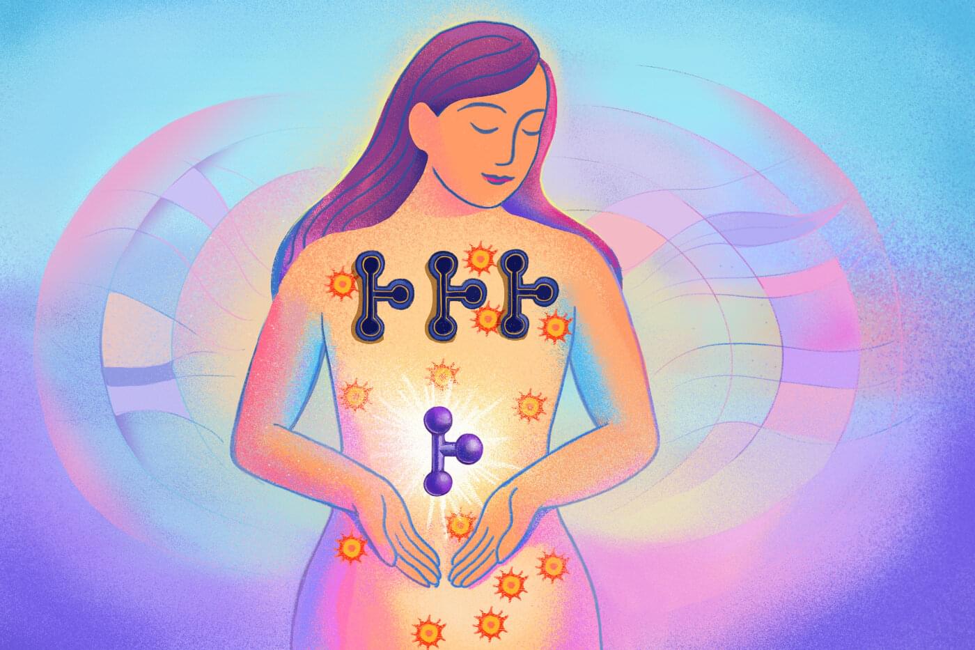
Northeastern University researchers have made a breakthrough drug discovery, developing the first synthetic endogenous cannabinoid compound, with repercussions for new therapeutics from pain and inflammation to cancer.
Spyros P. Nikas, an associate research professor in Northeastern’s Center for Drug Discovery, says that the discovery hinges on the distinction between two different kinds of cannabinoid chemicals, endogenous and exogenous. Exogenous cannabinoids are those produced outside the human body, like THC or CBD, both derived from the cannabis plant and present in marijuana.
Our own bodies, however, are also producing cannabinoids all the time. Called endogenous cannabinoids —or just “endocannabinoids”—these chemicals “modulate a wide range of physiological and pathophysiological responses,” Nikas says, processes that include mood, inflammation and even neurodegenerative disorders like Alzheimer’s and Parkinson’s. The research is published in the Journal of Medicinal Chemistry.
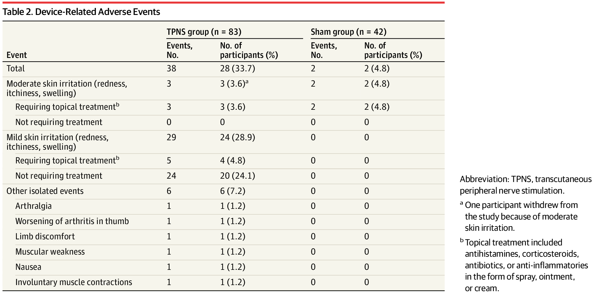
Essential tremor (ET), the most common upper limb tremor, can impair daily activities. In a multicenter RCT, an artificial intelligence–driven transcutaneous peripheral nerve stimulation (TPNS) device reduced mADL scores by 6.9 points at 90 days, compared with a 2.7-point reduction in the sham group.
Question Is an artificial intelligence (AI)–driven TPNS device superior to a sham device in reducing essential tremor?
Findings In this randomized clinical trial that included 125 adults with essential tremor, use of the TPNS device reduced the modified Activities of Daily Living score of the Essential Tremor Rating Assessment Scale by a clinically meaningful 6.9 points at 90 days, significantly more than the 2.7-point reduction seen in the sham-treated group.
Meaning The TPNS device improved activities related to upper limb tremor at 90 days and could be an effective noninvasive treatment for essential tremor.

Question What are the main predictors for high health care costs among patients with head and neck cancer?
Findings In this population-based cohort study, advanced cancer stage and receiving multiple treatment modalities were the strongest predictors of high health care costs. Female sex, older age, and lower socioeconomic status were associated with an increased likelihood for high health care costs, although with a weaker effect size.
Meaning Future research should focus on evaluating screening strategies and early diagnosis to assess their potential effects on cost reduction and improved outcomes for patients with head and neck cancer.
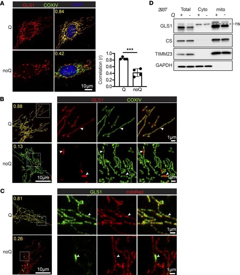
The team now report on a mechanism underlying kidney tumor cell growth involving the cancer-driving protein HIF2α that blocks clustering/activation of glutaminase—revealing a new therapeutic target in renal clear cell carcinoma:
The figure shows glutamine deprivation induces GLS1 clustering within mitochondria in HUVEC (top) and HELA (bottom) with mitochondrial protein (green) and GLS (red).
1Department of Medicine, Cardiovascular Institute, and Institute of Diabetes Obesity and Metabolism, and.
2The Abramson Family Cancer Research Institute, Department of Cell and Developmental Biology, Perelman School of Medicine, University of Pennsylvania, Philadelphia, Pennsylvania, USA.
3McAllister Heart Institute, School of Medicine, University of North Carolina, Chapel Hill, North Carolina, USA.

The incidence and prevalence of cutaneous melanoma in the US and worldwide have increased over the last 5 decades.
Cutaneous melanoma presents as a new, changing, or irregularly pigmented skin lesion. Risk factors for cutaneous melanoma include UV radiation exposure, skin type, presence of benign and atypical nevi, and personal or family history of melanoma.
This Review summarizes current evidence regarding the epidemiology, pathophysiology, diagnosis, and treatment of cutaneous melanoma.
Improvements in melanoma mortality over the last decade are attributed to the advent of multiple effective therapies,6 including immune checkpoint blockade with anti–cytotoxic T-lymphocyte–associated protein 4 (CTLA-4) antibodies (ipilimumab), anti–programmed cell death protein 1 (PD-1) antibodies (nivolumab, pembrolizumab), and anti–lymphocyte activation gene 3 protein (LAG-3) antibodies (relatlimab), as well as oral combination targeted therapy with B-Raf protein (BRAF) and mitogen-activated extracellular signal-regulated kinase (MEK) inhibitors (eg, encorafenib + binimetinib, vemurafenib + cobimetinib, dabrafenib + trametinib).
This review summarizes current evidence regarding epidemiology and risk factors, clinical presentation, diagnosis, and management of cutaneous melanoma (Box).
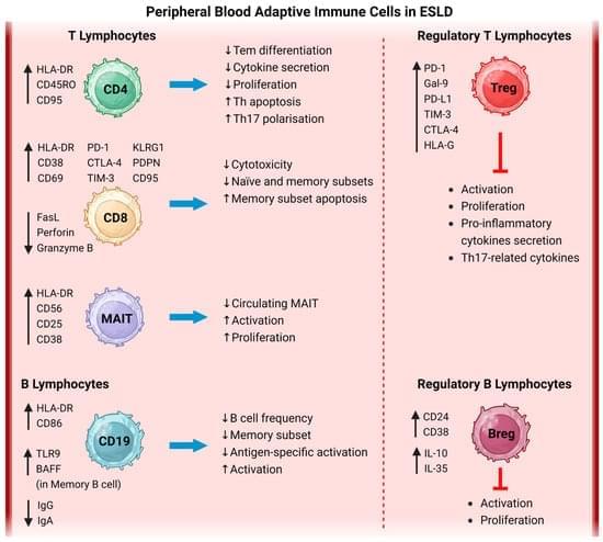
End-stage liver disease (ESLD) from acute liver failure to compensated advanced chronic liver disease and decompensated cirrhosis at different stages (chronic decompensation, acute decompensation with or without acute-on-chronic liver failure) has high disease severity and poor patient outcome. Infection is a common complication in patients with ESLD and it is associated with a high mortality rate. Multiple mechanisms are involved in this marked susceptibility to infections, noticeably the inadequate immune response known as immune paresis, as part of cirrhosis-associated immune dysfunction (CAID). Specifically in the adaptive immune arm, lymphocyte impairments—including inadequate activation, reduced ability to secrete effector molecules and enhanced immune suppressive phenotypes—result in compromised systemic immune responses and increased risk of infections.
