Formalin-fixed and paraffin-embedded (FFPE) tissue processing remains the gold standard for histopathological examination, enabling diagnosis of a variety of diseases, including cancers, infections, and many other abnormalities [1– 3]. FFPE relies on fixing tissues, embedding in paraffin wax, preparing thin tissue sections mounted on glass slides, followed by staining [e.g., hematoxylin and eosin (H&E)] to reveal tissue morphology (and in some cases, molecular composition) with high contrast. While these techniques provide high diagnostic utility and have been a mainstay tool in clinical pathology, they are time-and labor-intensive, requiring specialized equipment, trained personnel, and several chemical processing steps. The workflow, from sample acquisition to pathological evaluation, can take many hours to days [3– 5], limiting intraoperative or point-of-care decision-making, which ultimately delays patient care. Although frozen sections provide a faster alternative to FFPE sections, with turnaround times of 20 to 30 min, their diagnostic quality is substantially lower [3, 6, 7]. Furthermore, these analyses cannot be performed in low-resource settings without access to histology laboratories and the required equipment and supplies.
Slide-free histology has recently emerged as a promising alternative, offering the potential for direct point-of-care imaging of intact bulk tissues with no or minimal processing. This can enable real-time histological evaluation, reducing time to diagnosis while preserving tissue for downstream analyses. Several optical techniques have been applied for slide-free histology, including point scanning methods such as confocal microscopy [8– 11] and nonlinear multiphoton microscopy [12 – 15], which require some tissue processing for fluorescence labeling. More advanced label-free methods have also been introduced, such as stimulated Raman scattering [16 – 20] and photoacoustic microscopy (PAM) [21 – 28]. These techniques provide 3-dimensional (3D) histological images of un-(or minimally) processed tissues, but their clinical translation/adoption is hampered by the complexity, expense, and relatively large footprint of the systems. Wide-field imaging methods, including microscopy with ultraviolet surface excitation (MUSE) [29], fiber optic microscopy with ultraviolet surface excitation (FUSE) [30], and structured illumination microscopy (SIM) [31,32], have also been applied for slide-free imaging. These methods use much simpler instrumentation (i.e., lower costs, complexity, and footprint) with spatial multiplexing capabilities that offer faster acquisition times. However, these methods also require fluorescence labeling, which necessitates some degree of sample preparation. While fluorescence labeling is not as onerous a process as the entire FFPE and staining pipeline, it does introduce other caveats, including variable uptake of the fluorescent agent (particularly problematic for fresh tissues) and signal degradation via photobleaching. (Typically, tissues have to be fixed for optimal dye uptake, which adds to the processing time.) Furthermore, the very need for the fluorescent compounds and other necessary reagents can be a substantial added expense when imaging large volumes of tissues. Therefore, it is clinically desirable to have an imaging modality that is not only slide-free, fast, compact, and low cost but also label-free.
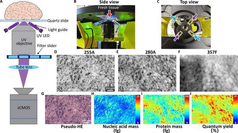
Deep-ultraviolet (UV) microscopy has recently emerged as a powerful approach for wide-field label-free molecular imaging [33 – 43]. By leveraging the intrinsic absorption properties of biomolecules, such as nucleic acids and proteins, in the deep-UV region of the spectrum (200 to 300 nm), this technique enables high-resolution, label-free, wide-field molecular imaging with simple and low-cost instrumentation. Deep-UV microscopy has been recently demonstrated for hematology [44] and histopathology applications, including label-free molecular analysis of prostate tissue sections [35,36]. One of the key advantages of deep-UV microscopy is its ability to enable quantitative mapping of biomolecules, providing pixel-level estimates of nucleic acid and protein mass with femtogram sensitivity derived directly from the absorption properties of these biomolecules [41,45]. By transforming qualitative images into quantitative maps, quantitative deep-UV microscopy has the potential to enable objective molecular phenotyping and real-time point-of-care assessment of the cellular and molecular composition, serving as an auxiliary readout for cytology and histology applications. However, prior implementations of deep-UV microscopy operate in transmission, hindering their applicability to unprocessed thick tissues.