Intraoperative functional brain mapping is an essential and intricate technique in modern-day glioma surgery. This article is not a review of the literature but of the technical protocol at our institution that has evolved over the recent decades to the current time and is intended to highlight details that enable us to perform maximal safe resection of gliomas.
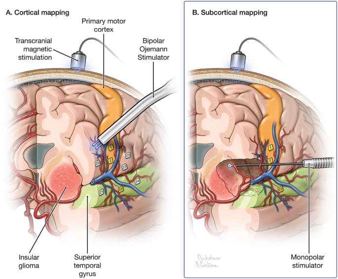
Prior to surgery, anatomical and functional imaging protocols are obtained to determine the tumor to be resected within its anatomical and functional environment. Preoperative assessments are used to determine which mapping procedures and tasks are most appropriate. Cortical and subcortical motor and language mapping using low and high frequency stimulation paradigms are applied when appropriate during resection. Methods to interpret findings and troubleshoot issues are reviewed herein.
All preoperative imaging including magnetic resonance imaging, magnetoencephalography of functional cortex, and diffusion tensor imaging of subcortical tracts are uploaded into the neuronavigation station and used throughout surgery for guidance. The decision to continue with tumor resection is based on constant feedback from the mapping paradigms as functional pathways are approached in real time. Both awake and asleep anesthesia regimens are utilized depending on the type of testing required to assess and preserve functional areas during tumor resection. Postoperatively, deficits are assessed using MRI along with clinical exam to predict whether they will be temporary or permanent.