Researchers at the Champalimaud Foundation in Lisbon have for the first time managed to identify with an imaging technique whether nervous impulses in the brain of rats are flowing in a “bottom-up” (feedforward), carrying information about visual input, or a “top-down” (feedback) direction, carrying information about expectations or predictions on a given task or about the perception of the world around us. Their results, published in Nature Communications, could have important implications for understanding changes in the brains of people with hallucinations, Alzheimer’s, schizophrenia, autism, and other conditions.
Joana Carvalho, first author of the new study, who at the time was working in the Preclinical MRI lab led by senior author Noam Shemesh (she has since become a group leader at Coimbra University), “came up with the ideas, did the experiments and analyzed the results. I just brought the MRI expertise,” says Shemesh good-humoredly. Co-author Koen V. Haak from Tilburg University (Netherlands) gave assistance with the computational models and the others helped with the experiments.
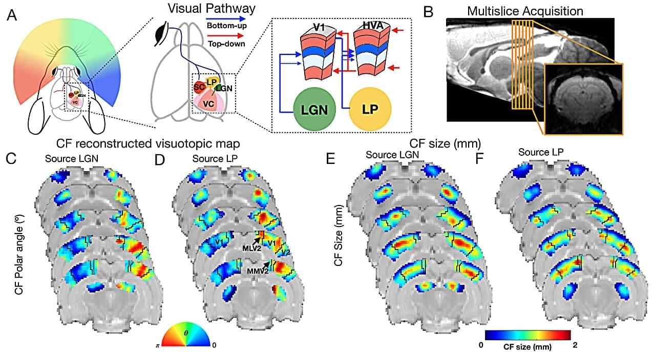
The team showed that spontaneous feedforward and feedback nervous impulses in these rodents (the brain never sleeps) each have a unique, distinct signature, which can be detected by using a method they developed, called uFLARE (UltraFast Layer-Resolved Encoding), a neuroimaging technique designed to map brain activity with unprecedented high temporal and spatial resolutions.