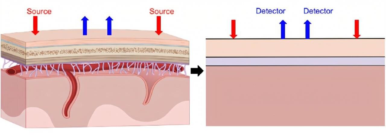
Near-infrared spectroscopy, or fNIRS, offers a way to monitor brain activity without surgery or radiation by tracking changes in blood flow and oxygenation. Light sources placed on the scalp send near-infrared light into the head, and detectors measure the light that scatters back. Because this light must pass through the scalp and skull before reaching the brain, the measured signal always includes a mix of superficial and cerebral contributions. Separating those signals has long been a central challenge for fNIRS researchers.
In a study published in Biophotonics Discovery, researchers from the Tufts University Diffuse Optical Imaging of Tissue Laboratory show that combining a specific source–detector geometry with a simple, anatomically informed tissue model can substantially improve how fNIRS data are interpreted.
By accounting for how light travels through layered head structures, the approach makes it possible to better isolate brain-specific signals without relying on complex imaging systems or subject-specific MRI scans.