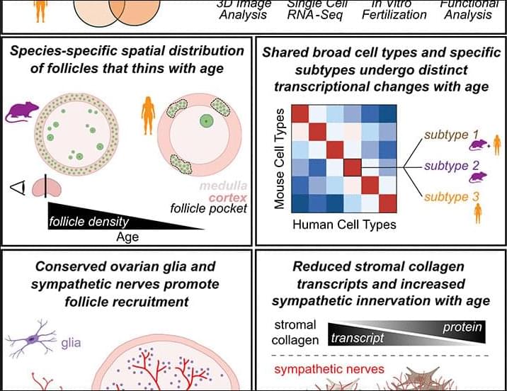
The mouse is a tractable model for human ovarian biology; however, its utility is limited by incomplete understanding of how transcription and signaling differ interspecifically and with age. We compared ovaries between species using three-dimensional imaging, single-cell transcriptomics, and functional studies. In mice, we mapped declining follicle numbers and oocyte competence during aging; in human ovaries, we identified cortical follicle pockets and decreases in density. Oocytes had species-specific gene expression patterns during growth that converged toward maturity. Age-related transcriptional changes were greater in oocytes than in granulosa cells across species, although mature oocytes change more in humans. We identified ovarian sympathetic nerves and glia; axon density increased in aged ovaries and, when ablated in mice, perturbed folliculogenesis.