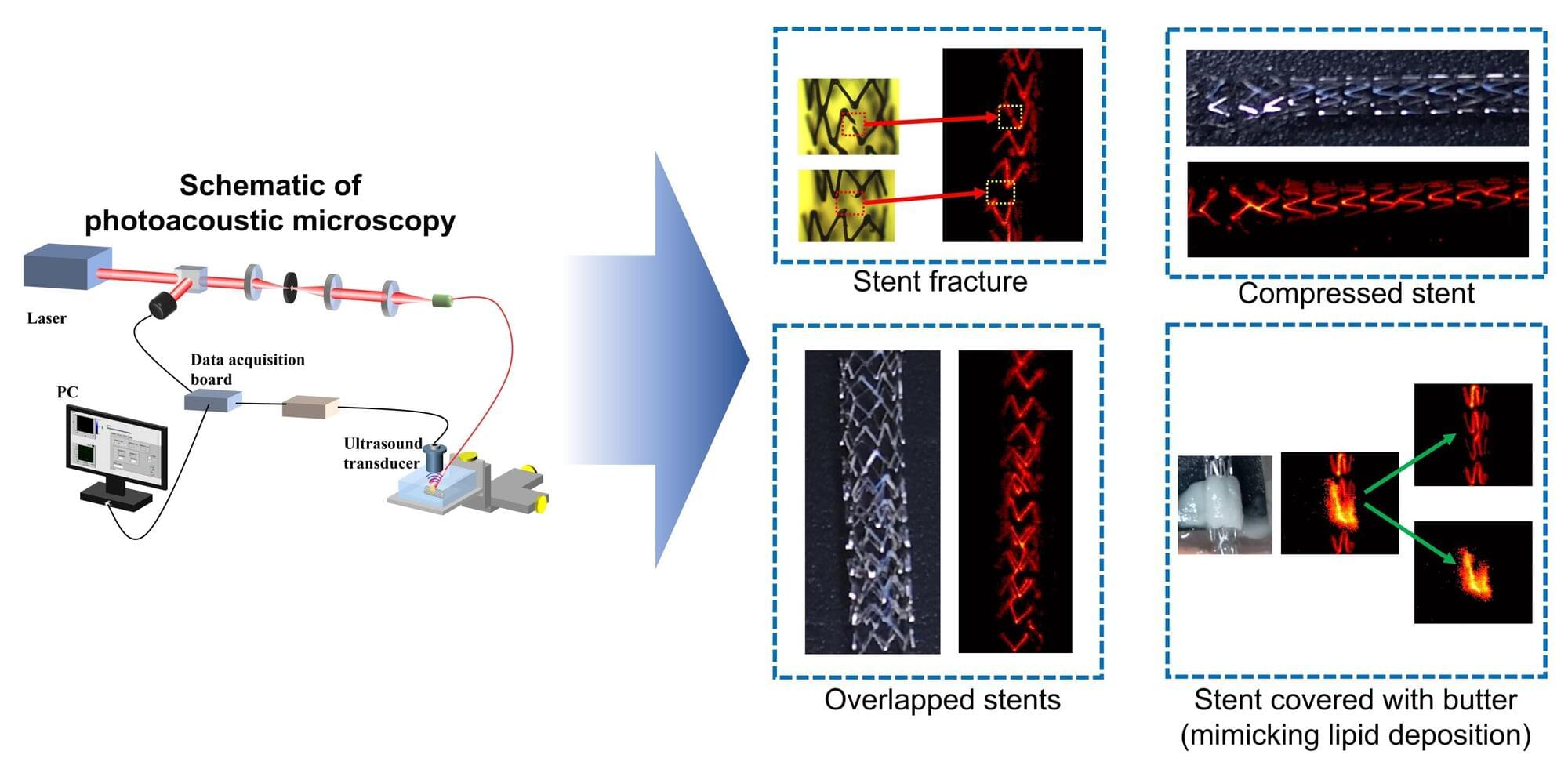
In a new study, researchers show, for the first time, that photoacoustic microscopy can image stents through skin, potentially offering a safer, easier way to monitor these life-saving devices. Each year, around 2 million people in the U.S. are implanted with a stent to improve blood flow in narrowed or blocked arteries.
“It is critical to monitor stents for problems such as fractures or improper positioning, but conventionally used techniques require invasive procedures or radiation exposure,” said co-lead researcher Myeongsu Seong from Xi’an Jiaotong-Liverpool University in China. “This inspired us to test the potential of using photoacoustic imaging for monitoring stents through the skin.”
In the journal Optics Letters, the researchers show that photoacoustic microscopy can be used to visualize stents covered with mouse skin under various clinically relevant conditions, including simulated damage and plaque buildup.
