Researchers from Aarhus University—in a major international collaboration—have developed a groundbreaking method that can provide more information from the tissue samples doctors take from patients every day.
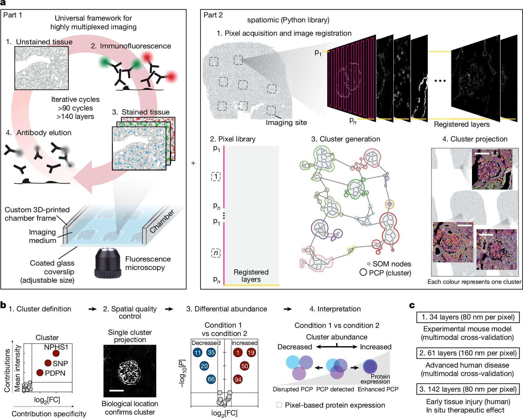
The new technique, called Pathology-oriented multiPlexing or PathoPlex, can look under a microscope at over 100 different proteins in the same small piece of tissue—instead of just 1–2 at a time, as is done now.
The technology, which has just been published in the journal Nature, combines advanced image processing with machine learning to map complex disease processes in detail.