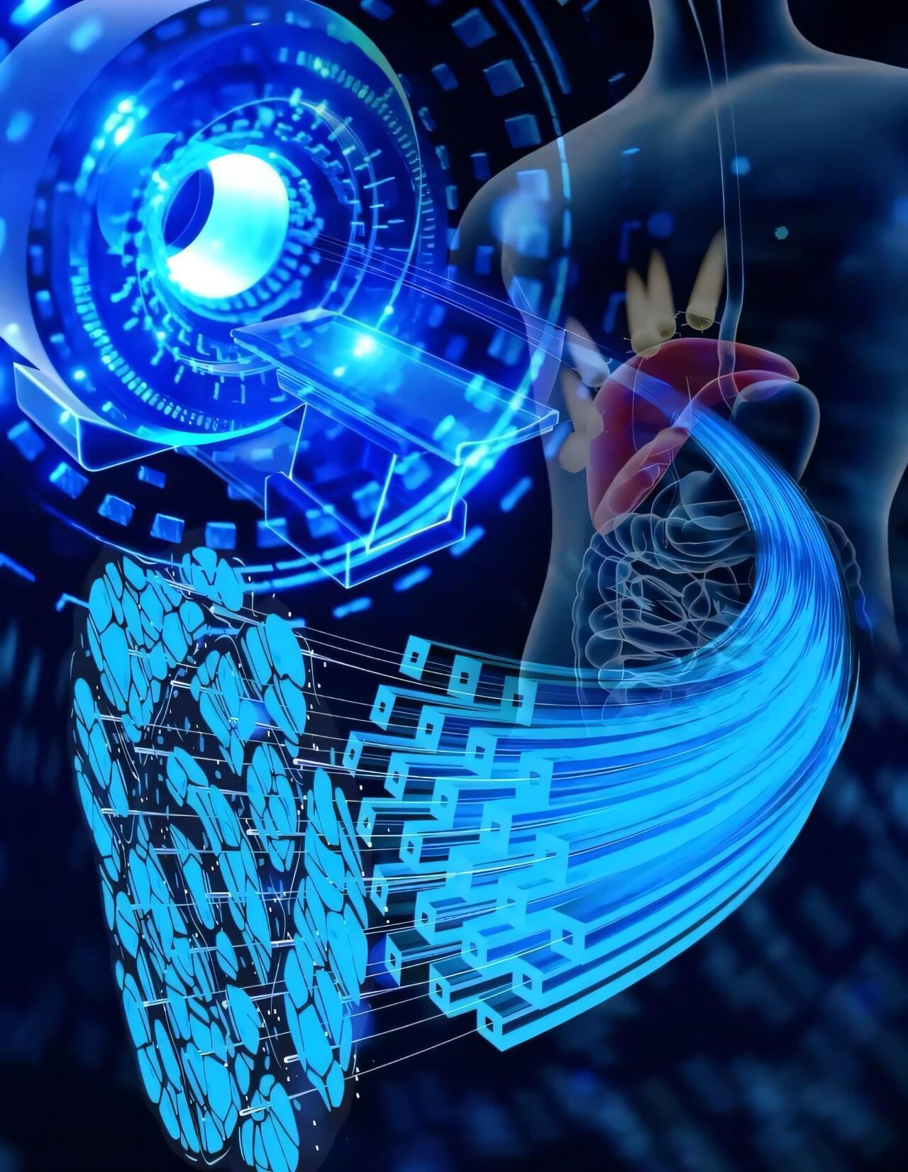
A research team has developed an innovative biomimetic dual-mode magnetic resonance imaging (MRI) nanoprobe for detecting early-stage liver fibrosis in non-alcoholic fatty liver disease (NAFLD).
The work, using the Steady High Magnetic Field Facility (SHMFF), was recently published in Advanced Science. The team was led by Prof. Wang Junfeng at the High Magnetic Field Laboratory, the Hefei Institutes of Physical Science of the Chinese Academy of Sciences.
NAFLD is a growing global health concern with even higher rates among individuals with obesity or type 2 diabetes. Detecting liver fibrosis early, before it becomes irreversible, is crucial for timely intervention and treatment. While MRI is a promising non-invasive tool for identifying liver fibrosis, traditional imaging techniques often lack the sensitivity to catch early-stage changes. Conventional contrast agents either face safety concerns or fail to target fibrotic tissues specifically.
