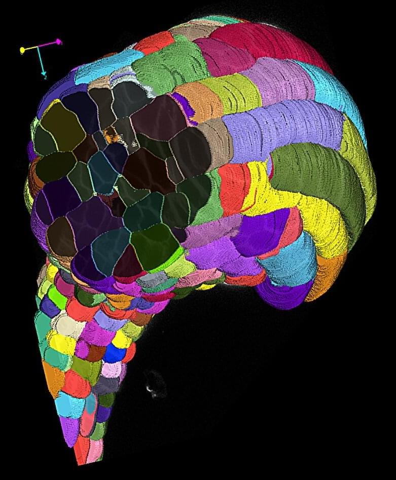
Identifying and delineating cell structures in microscopy images is crucial for understanding the complex processes of life. This task is called “segmentation” and it enables a range of applications, such as analyzing the reaction of cells to drug treatments, or comparing cell structures in different genotypes.
It was already possible to carry out automatic segmentation of those biological structures, but the dedicated methods only worked in specific conditions and adapting them to new conditions was costly. An international research team led by Göttingen University has now developed a method for retraining the existing AI-based software Segment Anything on over 17,000 microscopy images with over 2 million structures annotated by hand.
The new model is called Segment Anything for Microscopy and it can precisely segment images of tissues, cells and similar structures in a wide range of settings. To make it available to researchers and medical doctors, they have also created μSAM, user-friendly software to “segment anything” in microscopy images. The work is published in Nature Methods.