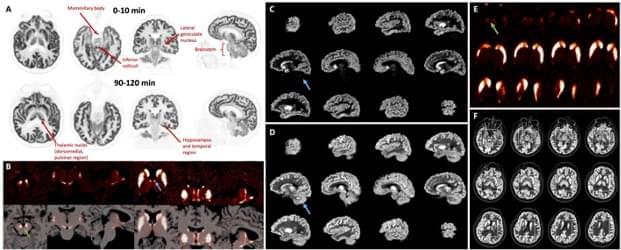
Toronto, Ontario —A new ultra-high-performance brain PET system allows for the direct measurement of brain nuclei as never before seen or quantified. With its ultra-high sensitivity and resolution, the NeuroEXPLORER provides exceptional brain PET images and has the potential to spur advances in the treatment of many brain diseases. This research was presented at the 2024 Society of Nuclear Medicine and Molecular Imaging (SNMMI) Annual Meeting, and the grouping of images highlighting targeted tracer uptake in specific brain nuclei has been selected as the 2024 SNMMI Henry N. Wagner, Jr., Image of the Year.
Each year, SNMMI chooses an image that best exemplifies the most promising advances in the field of nuclear medicine and molecular imaging. The state-of-the-art technologies captured in these images demonstrate the capacity to improve patient care by detecting disease, aiding diagnosis, improving clinical confidence, and providing a means of selecting appropriate treatments. This year, the SNMMI Image of the Year was chosen from more than 1,500 abstracts submitted for the meeting.
The image quality of PET systems has improved in recent years, mostly by increases in sensitivity, including enhanced time-of-flight capabilities. However, these systems have shown only minimal improvement in intrinsic resolution. To address these issues, researchers designed the NeuroEXPLORER PET scanner with a focus on ultra-high sensitivity and resolution, as well as continuous head motion correction.