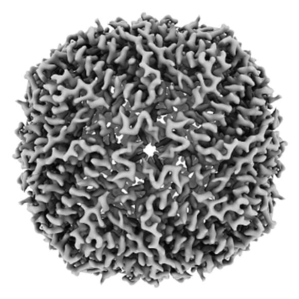
Talk to any structural biologist, and they’ll tell you how a cool new method is taking over their field. By flash freezing proteins and bombarding them with electrons, cryo–electron microscopy (cryo-EM) can map protein shapes with near-atomic resolution, offering clues to their function and revealing bumps and valleys that drug developers can target. The technique can catch wriggly proteins in multiple configurations, and it can even capture those that have been off-limits to traditional x-ray analysis because they stubbornly resist being crystallized. Many researchers expect cryo-EM will surpass x-ray crystallography in the number of new protein structures solved next year.
Yet for all its charms, cryo-EM has flaws: The freezing process is finicky, and the microscopes are expensive. High-end machines can cost more than $5 million to buy, about as much to install, and hundreds of thousands per year to operate and maintain. Many U.S. states—and countries—don’t have a single cryo-EM microscope. “The haves and have-nots is what it is right now,” says Rakhi Rajan, a structural biologist at the University of Oklahoma, which currently lacks one.
Researchers at the Medical Research Council’s Laboratory of Molecular Biology (LMB) have been working to democratize the field. Today, in the, the U.K. team describes cobbling together a prototype cryo-EM microscope that has solved its first structures. The machine—what LMB physicist Chris Russo calls a “cheap little hatchback” rather than a “Ferrari”—could rival high-end machines in capabilities for one-tenth of the cost.