“It is not enough to study brain connectivity with one single method, or even two,” says HBP Scientific Director and author of the Science article Katrin Amunts, who leads the Institute of Neuroscience and Medicine (INM-1) at Forschungszentrum Jülich and the C. & O. Vogt Institute of Brain Research at the University Hospital Düsseldorf. “The connectome is nested at multiple levels. To understand its structure, we need to look at several spatial scales at once by combining different experimental methods in a multi-scale approach and by integrating the obtained data into multilevel atlases such as the Julich Brain Atlas that we have developed.”
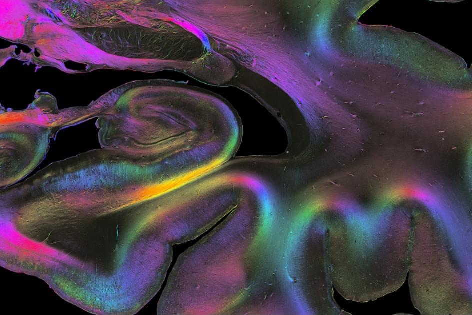
Markus Axer from Forschungszentrum Jülich and the Physics Department of the University of Wuppertal, who is the first author of the Science article, has together with his team at INM-1 developed a unique method called 3D Polarised Light Imaging (3D-PLI) to visualise nerve fibres at microscopic resolution. They trace the three-dimensional courses of fibres across serial brain sections with the aim of developing a 3D fibre atlas of the entire human brain.
Together with other HBP researchers from Neurospin in France and the University of Florence in Italy, Axer and his team have recently imaged the same tissue block from a human hippocampus using several different methods: anatomical and diffusion magnetic resonance imaging (aMRI and dMRI), two-photon fluorescence microscopy (TPFM) and 3D-PLI, respectively.