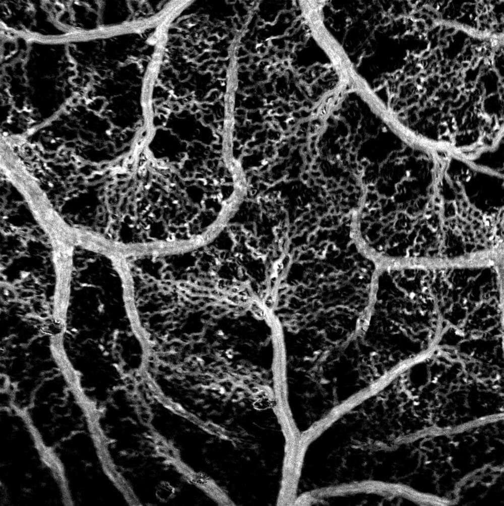
Researchers from the Skolkovo Institute of Science and Technology and Saratov State University have come up with an inexpensive method for visualizing blood flow in the brain. The new technique is so precise it discerns the motions of individual red blood cells — all without the use of toxic dyeing agents or expensive genetic engineering. The study was published in The European Physical Journal Plus.
To understand more about how the brain’s blood supply works, researchers map its blood vessel networks. The resulting visualizations can rely on a variety of methods. One highly precise technique involves injecting fluorescent dyes into the blood flow and detecting the infrared light they emit. The problem with dyes is they are toxic and also may distort mapping results by affecting the vessels. Alternatively, researchers employ genetically modified animals, whose interior lining of blood vessels is engineered to give off light with no foreign substances involved. Both methods are very expensive, though.
Researchers from Skoltech and Saratov State University have devised an inexpensive method for visualizing even the smallest capillaries in the brain. The method — which integrates optical microscopy and image processing — is dye-free and very fine-grained, owing to its ability to detect each and every red blood cell travelling along a blood vessel. Since the number of RBCs in capillaries is not that high, every cell counts, so this is an important advantage over other methods, including dye-free ones.