The extraordinary progress that has been achieved in the biomedical sciences in the modern era can be attributed in large measure to imaging technologies that have allowed scientists to observe the structure and function of tissues and organs in the context of their natural tissue environments in greater detail than possible with the naked eye.
But that ability has been limited to a handful of traditional animal model systems, including worms, flies and mice, either by tissue characteristics that make them amenable to imaging (such as a lack of natural pigmentation), or by the fact that the techniques used for preparing microscopic specimens in these models are not broadly applicable to a diverse range animal species.
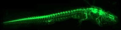
The development of a new imaging technique by Prayag Murawala, Ph.D., of the MDI Biological Laboratory and his collaborators, however, enables unprecedented insight subcellular structures, tissues, organs and even whole organisms, and—because of its wide applicability—broadens the range of animal models that can be studied, processes that can be explored and biological questions that can be addressed.