To tackle this problem, researchers at the RIKEN Center for Biosystems Dynamics Research identified a gel that closely mimics the physicochemical properties of organs that have undergone the tissue clearing process. Starting with computer simulations and following up with laboratory tests, the team optimized the soaking solution temperature, dye and antibody concentrations, chemical additives, and electrical properties to produce the best staining and imaging results. They then tested their method with more than two dozen commonly used dyes and antibodies on mouse and marmoset brains.
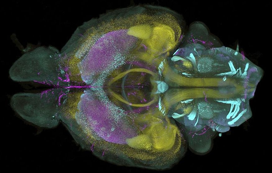
Scans of an entire mouse brain and one hemisphere of a marmoset brain—rendered into 3D using light sheet microscopy—revealed the similarity between the two animals’ neural vascular systems, showing the use of the system for comparative anatomy, the researchers report this week in. They also showed that they could simultaneously stain and image up to four molecular targets in a mouse brain, a feat that “has never been reported before,” says Ludovico Silvestri, of the European Laboratory for Non-linear Spectroscopy, who was not involved in the research.
The team also used its technique to image an entire infant marmoset and a small human brain sample—something that could one day lead to new understandings of solid tumors and neurodegenerative diseases. The team says its approach to optimizing staining can be applied to other techniques to advance the entire field of 3D imaging.