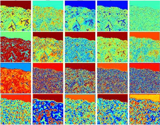
Tissue biopsy slides stained using hematoxylin and eosin (H&E) dyes are a cornerstone of histopathology, especially for pathologists needing to diagnose and determine the stage of cancers. A research team led by MIT scientists at the Media Lab, in collaboration with clinicians at Stanford University School of Medicine and Harvard Medical School, now shows that digital scans of these biopsy slides can be stained computationally, using deep learning algorithms trained on data from physically dyed slides.
Pathologists who examined the computationally stained H&E slide images in a blind study could not tell them apart from traditionally stained slides while using them to accurately identify and grade prostate cancers. What’s more, the slides could also be computationally “de-stained” in a way that resets them to an original state for use in future studies, the researchers conclude in their May 20 study published in JAMA Network Open.
This process of computational digital staining and de-staining preserves small amounts of tissue biopsied from cancer patients and allows researchers and clinicians to analyze slides for multiple kinds of diagnostic and prognostic tests, without needing to extract additional tissue sections.