To elicit VEPs, the eyelid corresponding to the stimulated retina was retracted temporarily while periodic 50 ms flashes were generated at 1 Hz from an array of white light-emitting diodes (LEDs). Neural response waveforms were temporally aligned to the stimulus onset. VEPs were calculated as the time-aligned averaged signals over 150 trials.
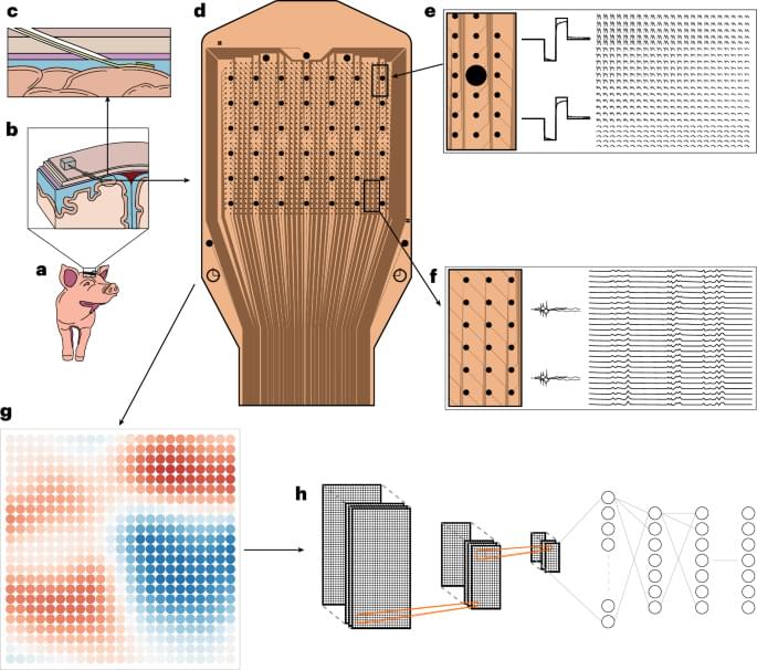
Electrical stimulation at the cortical surface was applied at one of the 200 µm electrodes, controlled by the Intan Technologies RHS controller and RHX software. Charge-balanced, biphasic, cathodic-first, 200 µs pulses of 100 µA peak current were delivered at 0.25 Hz. The evoked potentials were recorded over a series of trials. During analysis, for each trial and electrode, the Hjorth ‘activity’ of each trial was computed as the variance of the signal from 200 ms to 2,000 ms post-stimulation, and the average activity was taken over 40 trials.
A 1,024-channel array was placed over the sensorimotor cortex on each hemisphere following carefully sized bilateral craniectomies. Two Intan 1,024-channel RHD controllers were used to record from both arrays simultaneously.