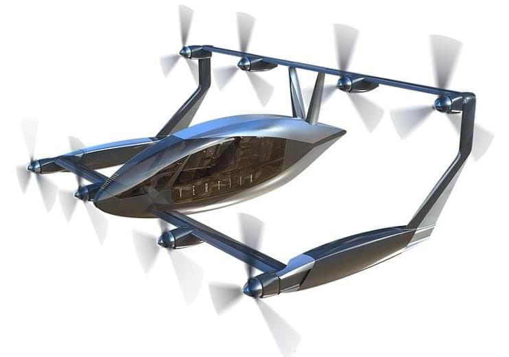
Toggle light / dark theme

A University of California, Berkeley professor stands at the front of the room, delivering her invited talk about the potential of genetic engineering. Her audience, full of organic farming advocates, listens uneasily. She notices a man get up from his seat and move toward the front of the room. Confused, the speaker pauses mid-sentence as she watches him bend over, reach for the power cord, and unplug the projector. The room darkens and silence falls. So much for listening to the ideas of others.

Many organic advocates claim that genetically engineered crops are harmful to human health, the environment, and the farmers who work with them. Biotechnology advocates fire back that genetically engineered crops are safe, reduce insecticide use, and allow farmers in developing countries to produce enough food to feed themselves and their families.

Now, sides are being chosen about whether the new gene editing technology, CRISPR, is really just “GMO 2.0” or a helpful new tool to speed up the plant breeding process. In July, the European Union’s Court of Justice ruled that crops made with CRISPR will be classified as genetically engineered. In the United States, meanwhile, the regulatory system is drawing distinctions between genetic engineering and specific uses of genome editing.

Nanolive, a spinoff company of École polytechnique fédérale de Lausanne (EPFL) in Switzerland, has just introduced a holographic microscope that can image live cells at high resolution over extended time periods.

Nanolive’s CX-A device relies on a low energy light beam to penetrate the sample, which does not interfere with internal cell activity. At every exposure, the system creates a 3D dataset of the sample, down to resolutions below 200 nanometers, which it can do repeatedly for hours at a time. Since entire 96-well plates can be imaged by the microscope, 96 individual experiments can be performed at once.

The system requires no cell preparation such as staining and doesn’t cause any phototoxicity or photo-bleaching in the samples.

The procedures implement national-level and DOD policies to protect information from foreign intelligence collection. It requires that the application of TEMPEST countermeasures be proportional and appropriate to the threat and potential damage to national security. It explains the selection, training, utilization, and operational requirements for appointment of an Army certified TEMPEST Technical Authority (CTTA) and provides Army protected distribution policy.

TEMPEST is a U.S. National Security Agency specification and a NATO certification referring to spying on information systems through leaking emanations, including unintentional radio or electrical signals, sounds, and vibrations.

The traditional approach for TEMPEST product approval provides for government supervision of evaluations to include testing oversight and technical reviews of both the TEMPEST test plans and test reports produced by a nation’s TEMPEST evaluation personnel.

NATO agreed on a scheme in 1981 to have vendors offer approved TEMPEST products for sale to NATO and NATO member nations.

Carl Malamud is on a crusade to liberate information locked up behind paywalls — and his campaigns have scored many victories. He has spent decades publishing copyrighted legal documents, from building codes to court records, and then arguing that such texts represent public-domain law that ought to be available to any citizen online. Sometimes, he has won those arguments in court. Now, the 60-year-old American technologist is turning his sights on a new objective: freeing paywalled scientific literature. And he thinks he has a legal way to do it.


A giant data store quietly being built in India could free vast swathes of science for computer analysis — but is it legal?

Scientists have produced what looks to be the most detailed magnetic resonance imaging (MRI) scan ever taken of the human brain anatomy, and are sharing their data with the public.

Thanks to an anonymous deceased patient whose brain was donated to science – and a gargantuan 100 hours of scanning with one of the most advanced MRI machines – the world now has an unprecedented view of the structures that make thought itself possible.

In a new study led by neuroimaging scientist Brian L. Edlow from Massachusetts General Hospital, researchers describe how they recorded their ultra-high resolution MRI dataset of the ex vivo specimen, offering a never-before-seen view of the “three-dimensional neuroanatomy of the human brain”.