Toggle light / dark theme

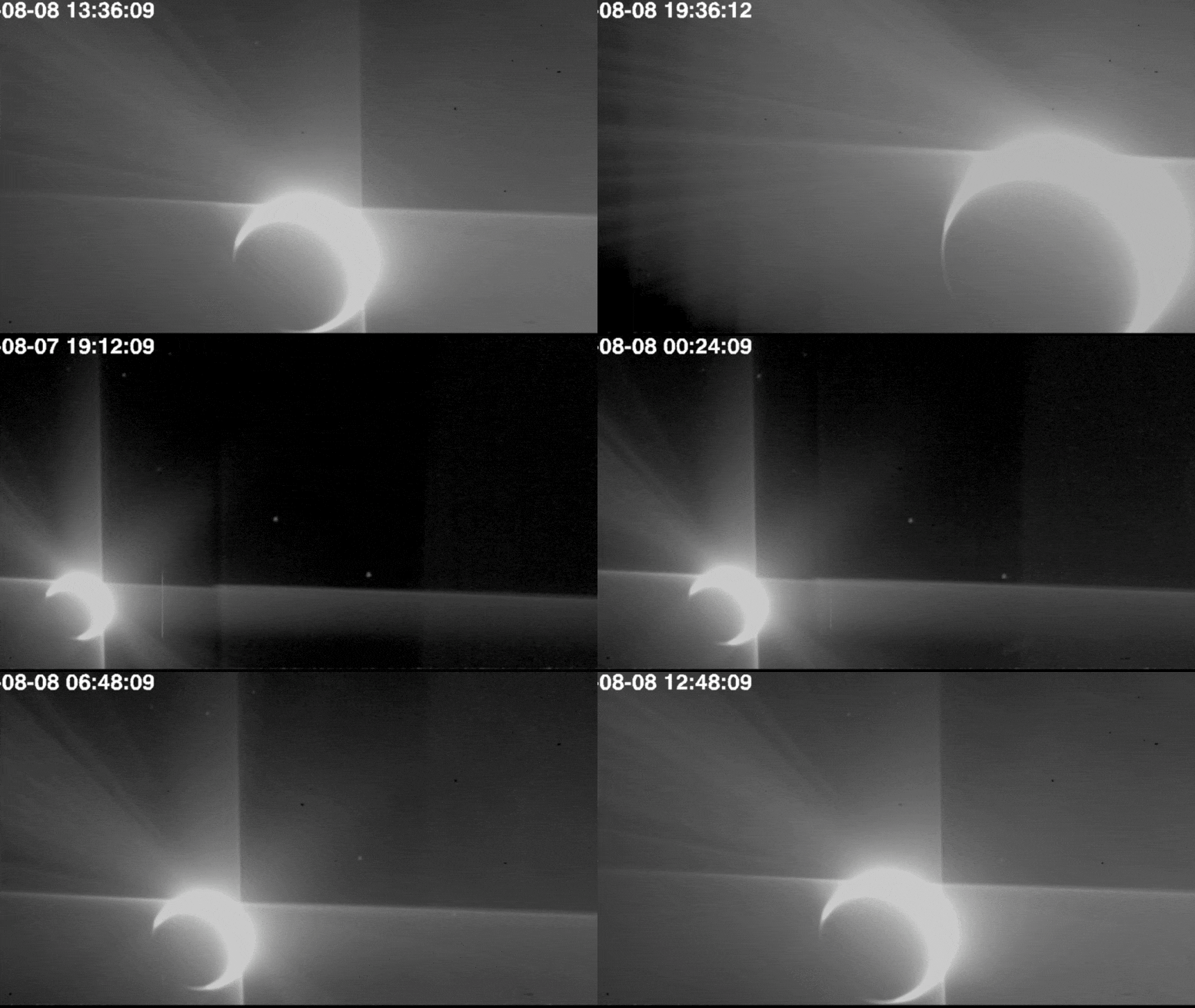
SpaceX has at long last transported its fifth full-scale Starship prototype to a nearby launch pad, kicking off a week of busy pre-test preparations while the sixth rocket is already nipping at its heels.

Starship SN5’s move to the pad ends the longest gap between full-scale prototype testing since Starship Mk1 was destroyed in November 2019, a partially-expected failure that began a more than three-month period of infrastructure upgrades. The first upgraded Starship (SN1) rolled to the pad on February 25th, followed by SN3 on March 29th and SN4 on April 23rd, indicative of a fairly consistent monthly cadence of Starship production (and destruction).

Almost exactly two months after its predecessor headed from the factory to SpaceX’s Boca Chica, Texas launch facilities, Starship SN5 has taken its place on a brand new launch mount. The cause of that month-long delay is fairly simple. When Starship SN4 exploded on May 29th, it damaged the existing launch mount and some additional ground support equipment (GSE), forcing SpaceX to scrap the destroyed mount and build a new one from scratch. In a matter of weeks, SpaceX’s crack team of pad engineers and technicians have done exactly that.

While some cholesterol is a healthy thing for properly functioning cells, too much of it can cause blockages in the arteries and heart trouble, along with a host of other negative health outcomes. Scientists have discovered a new mechanism by which a “bad” type of cholesterol gains entry to the cells, identifying a pair of proteins that work like an entry tunnel. These proteins show promise as new targets for drugs that could lower cholesterol levels in the blood to help prevent disease.

The discovery made by an international team of researchers was only possible thanks to advances in imaging technology that enabled them to inspect proteins at a near-atomic level. In this case, the team were investigating the role two proteins, NPC1 and NPC2, play in transporting low-density lipoprotein (LDL) cholesterol, often referred to as “bad” cholesterol, into our cells.

“Before 2013 we often had to theorise about how membrane proteins worked and how they functioned, but now we can actually see them, and seeing is believing,” says study author Prof Rob Yang from the UNSW in Australia. “We were able to look at the NPC1 and NPC2 proteins and see exactly the role they play in transporting this LDL cholesterol into the cell.”

A scientist in California has taken steps toward a long-sought gamma ray laser by harnessing positronium bubbles in special liquid helium. Positronium is a volatile, short-lived atom that seems kind of like hydrogen but has a positron—an antiparticle considered opposite to an electron, sometimes even called an antielectron—instead of a proton.

Holding positronium in liquid helium extends its viable stability, a relationship that’s decades old: “Positronium’s long lifetime in liquid helium was first reported in 1957,” says the press release, which links to a paper by physicist Richard A. Ferrell about the “reduced pickoff” positronium experiences when it can form a bubble inside liquid helium.

‘Twisted’ layers of 2D materials produce photonic topological transition at ‘magic’ rotation angles.

Monash researchers are part of an international collaboration applying ‘twistronics’ concepts (the science of layering and twisting 2D materials to control their electrical properties) to manipulate the flow of light in extreme ways.

The findings, published today in the journal Nature, hold the promise for leapfrog advances in a variety of light-driven technologies, including nano-imaging devices; high-speed, low-energy optical computers; and biosensors.

NDSU researchers recently developed a new method of creating quantum dots made of silicon. Quantum dots, or nanocrystals, are tiny nanometer-scale pieces of semiconductor that emit light when their electrons are exposed to UV light. The most common application of quantum dots is in QLED displays. Through their use, digital displays have become brighter and much thinner, resulting in improvements to television and, potentially, cell-phone technology.

Because silicon is abundant and nontoxic, silicon have unique technological appeal. Silicon quantum dots are currently being used for applications such as windows that remain transparent while serving as active photovoltaic collectors of energy, and they hold promise in medicine where quantum dots are coated with organic molecules to create nontoxic fluorescent biomarkers.

While traditional methods for creating silicon quantum dots require such as silicon tetrahydride (silane) gas or , the NDSU team’s research uses a liquid form of silicon to make the tiny particles at room temperature using relatively benign components.

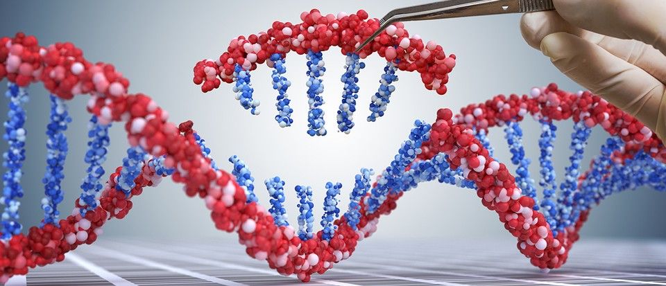
As part of their studies, the scientists also examined the mechanisms by which some of the modified drugs were altered by the cultured microbiomes. To understand exactly how the transformations occurred, they traced the source of the chemical transformations to particular bacterial species and to genes within those bacteria. They also showed that microbiome-derived metabolic reactions discoverable using their approach could be recapitulated in a mouse model, which is the first step in adapting the approach for human drug development.

The framework could feasibly be used to aid drug discovery by identifying potential drug-microbiome interactions early in development, and so inform on formulation changes. It could also be used during clinical trials to better analyze drug toxicity and efficacy, and be harnessed to help personalize treatment to the microbiome of each patient. This could help to predict how a certain drug will behave, and suggest changes to the therapeutic strategy if undesired effects are predicted. “Our framework identifies novel drug-microbiome interactions that vary between individuals and demonstrates how the gut microbiome might be used in drug development and personalized medicine,” the team concluded.

“This is a case where medicine and ecology collide,” said Jaime Lopez, a graduate student in the Lewis-Sigler Institute for Integrative Genomics and a co-first author on the study, who contributed the computational and quantitative analysis of the data. “The bacteria in these microbial communities help each other survive, and they influence each other’s enzymatic profiles. This is something you would never capture if you didn’t study it in a community.”


Researchers at Princeton University have developed a way of systematically evaluating how the microbial communities in our intestines can chemically transform, or metabolize, drugs that are taken orally, in ways that impact on their efficacy and potentially safety. The new methodology—which the team used to evaluate the gut microbiome’s effect on hundreds of common medications already on the market—provides a more complete picture of how gut bacteria metabolize drugs. The framework could also feasibly help in the development of drugs that are more effective, have fewer side effects, and are personalized to an individual’s microbiome.