We use cookies on our website. Some of them are essential, while others help us to improve this website and your experience. If you are under 16 and wish to give consent to optional services, you must ask your legal guardians for permission. We use cookies and other technologies on our website. Some of them are essential, while others help us to improve this website and your experience. Personal data may be processed (e.g. IP addresses), for example for personalized ads and content or ad and content measurement. You can find more information about the use of your data in our privacy policy. You can revoke or adjust your selection at any time under Settings.
Wearable technology is capable of tracking various measures of human health and is getting better all the time. New research shows how this could come to mean real-time feedback on posture and body mechanics. A research team at Cornell University has demonstrated this functionality in a novel camera system for the wrist, which it hopes to work into smartwatches of the future.
The system is dubbed BodyTrak and comes from the same lab behind a face-tracking wearable we looked at earlier in the year that is able to recreate facial expressions on a digital avatar through sonar. This time around, the group made use of a tiny dime-sized RGB camera and a customized AI to construct models of the entire body.
The camera is worn on the wrist and relays basic images of body parts in motion to a deep neural network, which had been trained to turn these snippets into virtual recreations of the body. This works in real time and fills in the blanks left by the camera’s images to construct 3D models of the body in 14 different poses.
The number of AI and, in particular, machine learning (ML) publications related to medical imaging has increased dramatically in recent years. A current PubMed search using the Mesh keywords “artificial intelligence” and “radiology” yielded 5,369 papers in 2021, more than five times the results found in 2011. ML models are constantly being developed to improve healthcare efficiency and outcomes, from classification to semantic segmentation, object detection, and image generation. Numerous published reports in diagnostic radiology, for example, indicate that ML models have the capability to perform as good as or even better than medical experts in specific tasks, such as anomaly detection and pathology screening.
It is thus undeniable that, when used correctly, AI can assist radiologists and drastically reduce their labor. Despite the growing interest in developing ML models for medical imaging, significant challenges can limit such models’ practical applications or even predispose them to substantial bias. Data scarcity and data imbalance are two of these challenges. On the one hand, medical imaging datasets are frequently much more minor than natural photograph datasets such as ImageNet, and pooling institutional datasets or making them public may be impossible due to patient privacy concerns. On the other hand, even the medical imaging datasets that data scientists have access to could be more balanced.
In other words, the volume of medical imaging data for patients with specific pathologies is significantly lower than for patients with common pathologies or healthy people. Using insufficiently large or imbalanced datasets to train or evaluate a machine learning model may result in systemic biases in model performance. Synthetic image generation is one of the primary strategies to combat data scarcity and data imbalance, in addition to the public release of deidentified medical imaging datasets and the endorsement of strategies such as federated learning, enabling machine learning (ML) model development on multi-institutional datasets without data sharing.
Tohid Didar and Jeff Weitz had a solution, but they also had a problem.
Didar, an associate professor of engineering and Weitz, a hematologist, professor of medicine and executive director of the Thrombosis & Atherosclerosis Research Institute, had collaborated to create a novel and highly promising material to improve the success of vascular grafts, but they needed a better way to test how well it worked.
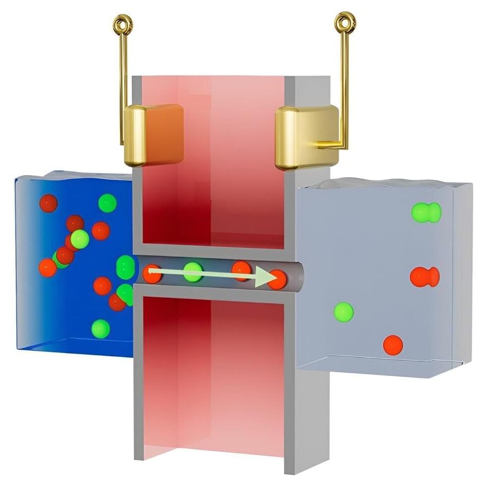
Their revolutionary idea was an engineered non-stick surface combined with biological components that can repel all but a targeted group of cells — those that form the natural lining of the body’s veins and arteries.
Sixteen people in six states have been infected with the listeria strain, resulting in one death and 13 hospitalizations, the agency said.
A tribute to the Garden City (Bengaluru), a ‘Walk in The Garden’ experience is designed to take passengers through green walls, hanging gardens, and outdoor gardens.
The cloud is everywhere, in almost every piece of technology we use. It powers all the ways we live and learn and play!
But what is the cloud? Imagine a massive network spanning the globe, with millions of computers all working to make our technology tick.
That network lives in hundreds of Microsoft datacenters around the world!
EXCERPT: ‘Prophets of Science Fiction’ hosted by Ridley Scott posits the science-fiction imaginings of the writers of the past are now becoming the science realities of our day. In this episode, Arthur C Clarke presents his 3 laws of prediction.
Making room for optimism.
2bsirius video about:
Arthur C. Clarke formulated the following three “laws” of prediction:
1. When a distinguished but elderly scientist states that something is possible, he is almost certainly right. When he states that something is impossible, he is very probably wrong.
2. The only way of discovering the limits of the possible is to venture a little way past them into the impossible.
3. Any sufficiently advanced technology is indistinguishable from magic.
For Shermer:
http://www.scientificamerican.com/article.cfm?id=shermers-last-law.
Full text of Shermers article:
For background on Arthur C. Clarke: