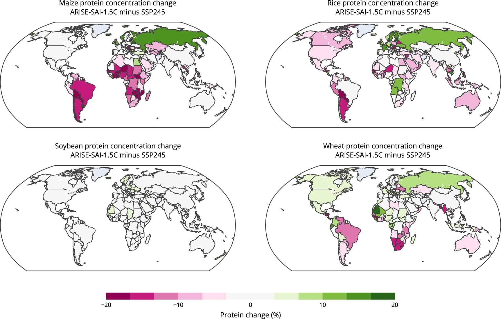
Attackers used 11 Go and 2 npm packages to spread malware across platforms, putting open-source developers at risk.

Researchers at European XFEL in Germany have tracked in real time the movement of individual atoms during a chemical reaction in the gas phase. Using extremely short X-ray flashes, they were able to observe the formation of an iodine molecule (I₂) after irradiating diiodomethane (CH₂I₂) molecules by infrared light, which involves breaking two bonds and forming a new one.
At the same time, they were able to distinguish this reaction from two other reaction pathways, namely the separation of a single iodine atom from the diiodomethane, or the excitation of bending vibrations in the bound molecule. The results, published in Nature Communications, provide new insights into fundamental reaction mechanisms that have so far been very difficult to distinguish experimentally.
So-called elimination reactions in which small molecules are formed from a larger molecule are central to many chemical processes—from atmospheric chemistry to catalyst research. However, the detailed mechanism of many reactions, in which several atoms break and re-form their bonds, often remains obscure. The reason: The processes take place in incredibly short times—in femtoseconds, or a few millionths of a billionth of a second.
Scientists at Johns Hopkins have grown a first-of-its-kind organoid mimicking an entire human brain, complete with rudimentary blood vessels and neural activity. This new “multi-region brain organoid” connects different brain parts, producing electrical signals and simulating early brain development. By watching these mini-brains evolve, researchers hope to uncover how conditions like autism or schizophrenia arise, and even test treatments in ways never before possible with animal models.
Newly developed DNA nanostructures can form flexible, fluid, and stimuli-responsive condensates without relying on chemical cross-linking, report researchers from the Institute of Science Tokyo and Chuo University, in the journal JACS Au.
Owing to a rigid tetrahedral motif that binds the linkers in a specific direction, the resulting string-like structures form condensates with exceptional fluidity and stability. These findings pave the way for adaptive soft materials with potential applications in drug delivery, artificial organelles, and bioengineering platforms.
Within living cells, certain biomolecules can organize themselves into specialized compartments called biomolecular condensates. These droplet-like structures play crucial roles in cellular functions, such as regulating gene expression and biochemical reactions; they essentially represent nature’s clever way of organizing cellular activity without the need for rigid membranes.
Both for research and medical purposes, researchers have spent decades pushing the limits of microscopy to produce ever deeper and sharper images of brain activity, not only in the cortex but also in regions underneath such as the hippocampus. In a new study, a team of MIT scientists and engineers demonstrates a new microscope system capable of peering exceptionally deep into brain tissues to detect the molecular activity of individual cells by using sound.
“The major advance here is to enable us to image deeper at single-cell resolution,” said neuroscientist Mriganka Sur, a corresponding author along with mechanical engineering Professor Peter So and principal research scientist Brian Anthony. Sur is the Paul and Lilah Newton Professor in The Picower Institute for Learning and Memory and the Department of Brain and Cognitive Sciences at MIT.
In the journal Light: Science and Applications, the team demonstrates that they could detect NAD℗H, a molecule tightly associated with cell metabolism in general, and electrical activity in neurons in particular, all the way through samples such as a 1.1 mm “cerebral organoid,” a 3D-mini brain-like tissue generated from human stem cells, and a 0.7 mm thick slice of mouse brain tissue.